Download
1 / 59
600 likes | 604 Views
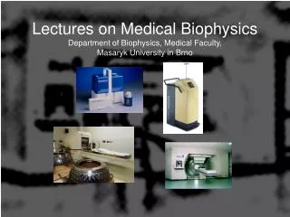
Lectures on Medical Biophysics Department of Biophysics, Medical Faculty, Masaryk University in Brno. Lectures on Medical Biophysics Department of Biophysics, Medical Faculty, Masaryk University in Brno. Nuclear medicine and radiotherapy. Nuclear medicine and radiotherapy.

E N D
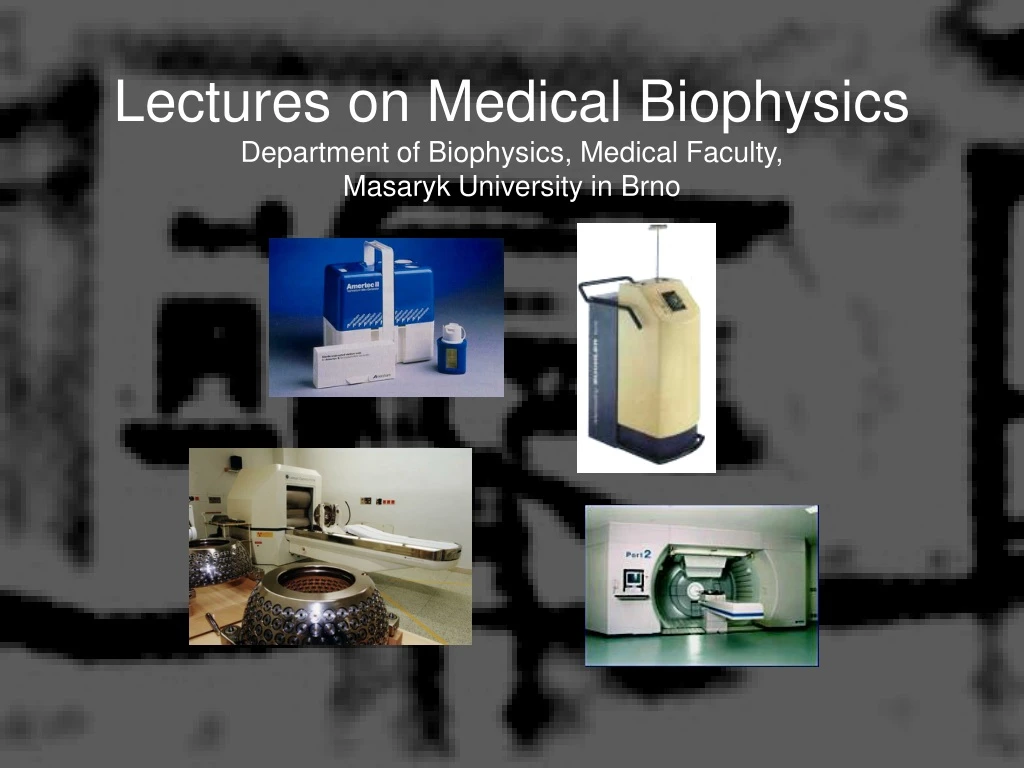
Lectures on Medical BiophysicsDepartment of Biophysics, Medical Faculty, Masaryk University in Brno
Lectures on Medical BiophysicsDepartment of Biophysics, Medical Faculty, Masaryk University in Brno Nuclear medicineand radiotherapy
Nuclear medicine and radiotherapy In this lecture we deal with selected methods of nuclear medicine and radiotherapy including their theoretical background: Radioactive decay Interactions of ionising radiation with matter Biological effects of ionising radiation Nuclear medicine • Tracing • Radioimmunoassay • Simple metabolic examinations • Imaging Radiotherapy • Sources of radiation – radioactive and non-radioactive • Methods of radiotherapy
Radioactivity • Radioactivity or radioactive decay is the spontaneous transformation of unstable nuclei into mostly stable nuclei. This is accompanied by the emission of gamma photons, electrons, positrons, neutrons, protons, deuterons and alpha particles. In some transformations, neutrinos and antineutrinos are produced. Unstable nuclei can be found naturally or created artificially by bombarding natural stable nuclei with e.g. protons or neutrons. • Radioactive decay has a stochastic character: it is not possible to determine which nucleus will decay at what time (tunnel effect).
Radioactive decay Laws valid for radioactive decay • Law of mass-energy conservation • Law of electric charge conservation • Law of nucleon number conservation • Law of momentum conservation
Radioactive decay Law of radioactive decay The activity A of a radioactive sample at a given time (i.e, the number of nuclei disintegrating per second, A = dN/dt) is proportional to the total number of undecayed nuclei present in the sample at the given time: • is the decay or transformation constant • Units of A are becquerel (Bq) [disintegrations per second, s-1] • (in the past:curie, 1 Ci = 3.7 x 10¹⁰ Bq) • The negative sign indicates that the number of undecayed nuclei is decreasing.
Radioactive decay This equation is solved by integration: Nt = N0.e-l.t A more useful equation for nuclear medicine and radiotherapy is (obtained by dividing the above equation by dt on both sides): At = A0.e-l.t A is activity
Radioactive decay Physical half-life • Tf – time in which the sample activity Atdecreases to one half of the initial value A0. Derivation: A0/2 = A0.e-l.Tf thus ½ = e-l. Tf • taking logarithm of both sides of the equation and rewriting: Tf = ln2/lf thus Tf = 0,693/lf
Radioactive decay Biological and effective half-life • Tb – biological half-life – time necessary for the physiological removal of half of a substance from the body • lb – biological constant – relative rate of a substance removal • Biological and physical processes take place simultaneously. Therefore, we can express the Tef – effective half-life and lef – effective decay constant • The following equations hold: lef = lb + lf and 1/Tef = 1/Tf + 1/Tb, thus
Radioactive decay Technetium generator During radioactive decay, a daughter radionuclide is produced. In cases when the half-life of the parent radionuclide is much longer than the half-life of the daughter radionuclide both parent and daughter end up with the same activity (radioactive equilibrium). An example of practical importance of the radioactive equilibrium in clinical practice – production of technetium for diagnostics: Mo-99 half-life is 99 hrs., Tc-99m half-life is 6 hrs. l1N1 = l2N2
Radioactive decay Classes of radioactive decay a (alpha) decay Seaborgium transforms in rutherfordium. Helium nucleus – a particle – is liberated. Daughter nucleus recoils as a consequence of the law of momentum conservation.(http://www2.slac.stanford.edu/vvc/theory/nuclearstability.html)
Radioactive decay Classes of radioactive decay b decay is an isobaric transformation in which besides the b particles are formed also neutrinos (electron antineutrino or electron neutrino ne) b (beta) decay = emission of an electron or positron K-capture
Radioactive decay Classes of radioactive decay • (gamma) decay Transformation of dysprosium nucleus in excited state • The other classes of radioactive decay: • Emission of proton, deuteron, neutron … • Fission of heavy nuclei
Interaction of ionising radiation with matter • The interaction of radiation with matter is usually accompanied bythe formation ofsecondary radiationwhich differs from the primary radiation by lower energy and often also by kind of particles. • Primary or secondary radiation directly or indirectlyionisesthe medium, and creates alsofree radicals. • A portion of the radiation energy is always transformed intoheat. • The energy loss of the particles of primary radiation is characterised by means of LET,linear energy transfer,i.e. energy loss of the particle in given medium per unit length of its trajectory. The higher the LET the more damaging is the radiation to tissues and the higher the risk from the radiation.
Interaction of ionising radiation with matter Attenuation of X / gamma radiation When a beam of X or gamma radiation passes through a substance: absorption + scattering = attenuation A small decrease of radiation intensity -dI in a thin substance layer is proportional to its thickness dx, intensity I of radiation falling on the layer, and a specific constant m: -dI = I.dx.m After rewriting: dI/I = -dx.m After integration: I = I0.e-m.x I is intensity of radiation passed through the layer of thickness x, I0 is the intensity of incident radiation, m islinear coefficient of attenuation[m-1] (depending on photon energy, atomic number of medium and its density).
Interaction of ionising radiation with matter Interactions of photon radiation (X-rays and gamma rays) • Photoelectric effect and Compton scattering – see the lecture on X-ray imaging. • Electron - positron pair production (PP) – very high energy photons only. The energy of the photon is transformed into mass and kinetic energy of an electron and positron. The mass-energy E in each particle is given by: E = m0 c2 (= 0,51 MeV), m0 is rest mass of an electron / positron (masses of electron and positron are equal), c is speed of light in vacuum. Energy of the photon must be higher than twice the energy calculated using the above formula (1.02 MeV). We can write: E = h.f = (m0.c2 + Ek1) + (m0.c2 + Ek2) • Terms in brackets: mass-energies of created particles, Ek1 a Ek2 kinetic energies of these particles. • The positron quickly interacts (annihilates) with any nearby electron, and two photons originate, each with energy of 0.51 MeV.
Interaction of ionising radiation with matter Electron - positron pair production
Interaction of ionising radiation with matter Interaction of corpuscular radiation with tissue • b radiation=fast electrons or positrons – ionise the medium as in X-ray production. Trajectory of a b particle is several millimetres in aqueous medium. • a radiationionises directly by impacts. There is formed big number of ions along its very short trajectory in medium (mm) – so it loses energy very quickly along a short trajectory (= very high LET) . • Neutronsionise by elastic and non-elastic impacts (scatter) with atomic nuclei. The result of anelastic scatterdiffers according to the ratio of neutron mass and atom nucleus mass. When afast neutronhits the nucleus of a heavy element, it bounces off almost without energy loss. Collisions with light nuclei lead to big energy losses. Innon-elastic scatter, the slow(moderated, thermal) neutronspenetrate into the nucleus, and if they are emitted from it again, they do not have the same energy like the incident neutrons. They can lead to the emission of other particles or fission of heavy nuclei.
Interaction of ionising radiation with matter Main quantities and units for measurement of ionising radiation • Absolute value of particle energy is very small. Therefore, theelectron volt(eV) was introduced. 1 eV is the kinetic energy of an electron accelerated from rest by electrostatic field of the potential difference 1 volt. 1 eV = 1.602×10⁻¹⁹ J. • Energy absorbed by the medium is described byabsorbed dose (D)- unitgray, Gy). It is the amount of energy absorbed per unit mass of tissue. Gray = J.kg-1 • Dose rateexpresses the absorbed dose in unit time [J.kg-1.s-1]. The same absorbed dose can be reached at different dose rates during different time intervals. • The radiation hazard to biological objects depends mainly on the absorbed dose and the type of radiation. The radiation weighting factor is a number which indicates how hazardous a type of radiation is (the higher the LET the higher the radiation weighting factor). • Equivalent dose Deis defined as the product of the absorbed dose and the radiation weighting factor. The unit of Equivalent dose is thesievert (Sv).
Biological effects of ionising radiation • Physical phase– time interval of primary effects. Energy of radiation is absorbed by atoms or molecules. Mean duration is about 10-16 s. • Physical-chemical phase– time interval of intermolecular interactions (energy transfers). About 10-10 s. • Chemical (biochemical) phase– free radicals are formed. They interact with important biomolecules, mainly with DNA and proteins. About 10-6 s. • Biological phase– a complex of interactions of chemical products on various levels of the living organism and their biological consequences. Depending on these levels, the duration ranges from seconds to years.
Biological effects of ionising radiation • Direct action (hits) – physical and physical-chemical process of radiation energy absorption, leading directly to changes in important cellular structures. It is the most important action mechanism in cells with low water content. Theory of direct action is called target theory. It is based on physical energy transfer. • Indirect effects are mediated by water radiolysis products, namely by free radicals H* a OH*. It is most important in cells with high water content. The free radicals have free unpaired electrons which cause their high chemical reactivity. They attack chemical bonds in biomolecules and degrade their structure. Theory of indirect action – radical theory – is based on chemical energy transfer.
Biological effects of ionising radiation Effects on the cell In proliferating cells we find these levels of radiation damage: • Transient stopping of proliferation • Reproductive death of cells(vital functions are maintained but proliferation ability is lost) • Instantaneous death of cells Cell sensitivity to ionising radiation (radiosensitivity), or their resistance (radioresistance) depends mainly on the repair ability of the cell.
Biological effects of ionising radiation Effects on the cell Factors influencing biological effects in general: • Physical and chemical: equivalent dose, dose rate, temperature, spatial distribution of absorbed dose, presence of water and oxygen. • Biological: species, organ or tissue, degree of cell differentiation, physiological state, spontaneous ability of repair, repopulation and regeneration. Sensitivity of cells is influenced by: • Cell cycle phase(S-phase!) • Differentiation degree. Differentiated cells are less sensitive. • Water and oxygen content. Direct proportionality (+,+) Very sensitive are e.g. embryonic, generative, epidermal, bone marrow and alsotumour cells
Biological effects of ionising radiation Tissue sensitivity Arranged according to the decreasing radiosensitivity: lymphatic spermatogenic epithelium of testis bone marrow gastrointestinal epithelium ovaries cells of skin cancer connective tissue liver pancreas kidneys nerve tissue brain muscle Typical symptoms of radiation sickness: 1. Non-lethal – damage to the erythropoiesis (bone marrow), effects on gonads 2. Lethal – gastrointestinal syndrome (damaged epithelium), skin burning, damage to suprarenal glands, damaged vision, nerve syndrome (nerve death) Late sequels – cumulative– genetic damage, cancer
Nuclear medicine Nuclear medicine • Tracing • Radioimmunoassay • Simple metabolic examinations • Imaging
Nuclear medicine Tracing and radioimmunoassay • Tracing: radionuclide is administered into body and its physiological fate is followed. Radioactivity is measured in body fluids or tissue samples. Compartment volumes – e.g. free water, blood, fat etc. – are often determined. We administer defined amount (known activity) of a radionuclide, and determine its concentration in taken samples after certain time. Then is possible to calculate what is the volume, in which the radionuclide is present. • Radioimmunoassay (RIA) is a method of clinical biochemistry and haematology. It is used for determination of low concentrated substances, e.g. hormones in blood. Radionuclide is applied outside the body and the antigen-antibody interaction is studied in vitro. The antigen is labelled by radionuclide. In RIA and tracing, mainly b-emitters are used (tritium, iodine-125, iron-59 etc.), because the detector can be very close to the radioactive sample. Both methods were very important but today seem obsolete.
Nuclear medicine Scintillation counter and scintigraphy(history of medicine) • Scintillation counter consisted of a scintillation detector, mechanical parts and a lead collimator. The collimator enabled the detection of radiation only from a narrow spatial angle, in which the examined body part was located. Signals of the detector were amplified, counted and recorded. • Scintigraphywas used mostly for examination of kidneys and thyroid gland – by means of gamma-emitters: iodine-131 or technetium-99m. Tc-99m has a short half-life (6 hours vs. 8 days in I-131). Technetium is prepared directly in dept. of nuclear medicine in technetium generators. • Iodine used for thyroid was administered as KI, for kidneys was used technetium-labelled DTPA (diethylen-triamin-penta-acetic acid). Tc-99m is almost an ideal diagnostic radionuclide – fastly excreted, short half-life, almost pure gamma rays. (Iodine-131 produces also b-particles which increases radiation dose without any benefit).
The Gamma Camera MCA photomultiplier tubes (now being replaced by a flat digital sensor) thin (about 1.5 cm) NaI phosphor crystal parallel hole Pb collimator for localisation
Nuclear medicine Gamma-camera • The digital sensor / photomultiplier signals carry information about the position of the scintillation events. However, a defined point on the crystal has to correspond with defined point of the examined body part – we obtain an image of radionuclide distribution in the body. This can be achieved only by collimators. • Anger cameras show the radionuclide distribution very quickly. Therefore they can be used for observation of fast processes, including blood flow in coronary arteries. They can also move along the body. Physiologic (functional) information is obtained or metastases found (if the radionuclide is entrapped there - iodine-131 or technetium-99m). A whole body scan showing metastases of a bone tumour
Nuclear medicine SPECT – single photon emission computed tomography • Photons of radiation are detected from various directions, which allows reconstruction of a cross-section. • Most frequent arrangements and movements of detectors: • Anger scintillation camera revolves around the body. • Many detectors are arranged around the body in a circle or square. The whole system can revolve around the body in a spiral (helix).
Nuclear medicine Principle of SPECT In SPECT, common sources of radiation (iodine-131, technetium-99m) are used. An object with a radiation source Z is surrounded by scintillation detectors F with collimators K. The collimators allow detection only of gamma-rays falling normally onto the detector blocks. It enables us to localise the source of rays.
Nuclear medicine SPECT – imageshttp://www.physics.ubc.ca/~mirg/home/tutorial/applications.html#heart Perfusion of heart in different planes. „Hot“ regions are well blood supplied parts of the heart Brain with „hot“ regions
Nuclear medicine PET - positron emission tomography • In PET, positron emitters are used. They are prepared in accelerators, and their half-lives are very short – max. hours. For that reason the examination must be done close to the accelerator, in a limited number of medical centres. • The positrons travel only very short distance, because they annihilate with electrons forming two gamma photons (0.51 MeV), which move in exactly opposite directions. These photons can be detected by two opposite detectors connected in a coincidence circuit. Voltage pulses are recorded and processed only when detected simultaneously in both detectors. Detectors scan and rotate around the patient's body. • The spatial resolution of PET is substantially higher than in SPECT. The positron emitters are attached to e.g. glucose derivatives, so that we can obtain also physiological (functional) information. PET of brain visualises those brain centres which are at the moment active (have increased uptake of glucose). PET allows to follow CNS activity on the level of brain centres.
Nuclear medicine PET principle Explanation of the high spatial resolution of PET: The opposite detectors in a coincidence circuit. A source of radiation Z is detected only when lying on a line connecting the detectors. Detector A but not the detector B can be hit through a collimator from the source Z2, because this source is outside the detection angle of B. In SPECT, the signal detected by A from Z1 would be partially overlapped by the signal coming from source Z2.
Nuclear medicine Functional PET of brainhttp://www.crump.ucla.edu/software/lpp/clinpetneuro/lggifs/n_petbrainfunc_2.html resting Music – a non-verbal acoustic stimulus Visual stimulus
Nuclear medicine intensive thinking remembering a picture skipping on left leg
Nuclear medicine Brain tumour - astrocytoma FDG – fluorodeoxyglucose, F-18
Radiotherapy • Sources of radiation – radioactive - non-radioactive • Methods of radiotherapy
Radiotherapy Sources of radiation - radioactive • Artificial radionuclides are used.The source is in direct contact with a tissue or is sealed in an envelope (open or closed sources). • The open sources: • (1) Can be applied by metabolic way. Therapy of thyroid gland tumours by radioactive iodine I-131, which is selectively captured by the thyroid. • (2) Infiltration of the tumour by radionuclide solution, e.g. a prostate tumour by the colloid gold Au-198. This way of application is seldom used today as well. • The closed sources are more widely used today: • (1) Needles with a small amount of radioactive substance. They usually contain cobalt Co-60 or caesium Cs-137. The needles are applied interstitially (directly into the tumour). • (2) The sources are also inserted into body cavities (intracavitary irradiation - afterloaders). • (3) Large irradiation devices (‘bombs’) for teletherapy. The radionuclide is enclosed in a shielded container. The radioactive material is moved into working position during irradiation. The most commonly used are cobalt Co-60 or caesium Cs-137. These devices are obsolete today.
Radiotherapy „The cobalt bomb“
Radiotherapy „The cobalt bomb“http://www.cs.nsw.gov.au/rpa/pet/RadTraining/
Radiotherapy Leksell Gamma Knife (still used) • 1951 – idea of radiosurgery by L. Leksell of Sweden • The Leksell Gamma Knife is used for treatment of some brain tumours and other lesions (aneurysms, epilepsy etc.) • 201 Co60 sources are placed in a central unit with diameter of 400 mm in 5 circles, which are separated by the angle of 7,5 deg. Each beam is collimated by a tungsten collimator with a conical channel and a circular orifice (4, 8, 14 a 18 mm in diameter). The focus is in the centre where all the channel axes (beams) intersect. The beams converge in the common focus with accuracy of 0.3 mm. • The treatment table is equipped by a movable couch. The patient‘s head is fastened in the collimator helmet. It is attached to the couch, which can move inside the irradiation area.
Radiotherapy Leksell Gamma Knife
Radiotherapy Leksell Gamma Knife • A Leksell stereotactic coordinate frame is attached to patient‘s head by means of four vertical supports and fixation screws. The head is so placed in a 3D coordinate system, where each point is defined by coordinates x, y, z. Their values can be read on the frame. The target area can be located with an accuracy better than ± 1 mm. • A radiological image of the lesion is transferred to the planning system which calculates the total dose from all the 201 sources. By connecting of points with the same dose a curve – isodose – is constructed. The borders of treated lesion should correspond with isodose showing 50-70% of dose maximum. The isodoses copy precisely the outlines of the pathologic lesion in tomographic scans.
Radiotherapy Leksell Gamma Knife
Radiotherapy Leksell Gamma Knife
Radiotherapy Afterloaderworks with Ir-192. An instrument for safe intracavitary irradiation. applicators Control unit main unit phantom
Radiotherapy Radiation sources – non-radioactive • X-ray tube devices: Therapeutic X-ray tubes differ in construction from diagnostic X-ray tubes. They have larger focus area, robust anode and effective cooling. They are (were) produced in three sorts: • low-voltage(40 - 100 kV) for contact surface therapy. The radiation is fully absorbed by a soft tissue layer 2 - 3 cm thick. e.g., Chaoul lamp. • medium-voltage (120 - 150 kV) for brachytherapy – from distance of max. 25cm. They were used to irradiate tumours at max depth 5 cm. • ortho-voltage (160 - 400 kV) for teletherapy (deep irradiation from distance). These have been replaced by the radionuclide sources and accelerators. B) Electron Accelerators: X-rays with photon energy above 1 MeV and g-radiation with photon energy above 0,66 MeV are used for megavoltage therapy. Their sources are mainly electron accelerators. The accelerated electrons are usually not used for direct irradiation but the production of high-energy X-rays.
Radiotherapy The linear accelerator CLINAC 2100C in Masaryk memorial institute of oncology in Brno
Radiotherapy The linear acceleratorhttp://www.cs.nsw.gov.au/rpa/pet/RadTraining/MedicalLinacs.htm