Download
1 / 37
520 likes | 1.27k Views
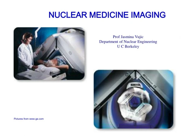
Nuclear Medicine Imaging. Overview. Nuclear medicine: Therapeutic and diagnostic use of radioactive substances Radioactivity: Naturally occurring radioisotopes (radioactive isotopes) discovered 1896 by Becquerel

E N D
Overview • Nuclear medicine:Therapeutic and diagnostic use of radioactive substances • Radioactivity: • Naturally occurring radioisotopes (radioactive isotopes) discovered 1896 by Becquerel • First artificial radioisotopes produced by the Curies 1934 (32P) “Radioactivity,” “Radioactive” • 1947 - Kohman: “Radionuclide” = nucleus of measurable half-life • 1935 - Hevesy uses 32P for metabolic studies with Geiger-Muller counter • 1949 - First radionuclide imaging by Cassen of 131I uptake in thyroid gland(scintillator+PMT, scanner, collimator,1/4” spatial resolution) • 1957 - Anger camera (planar imaging) • 1960 - Kuhl & Edwards construct Mark IV scanner (~10 years before x-ray CT) • 1977 – Kayes & Jaszczak develop SPECT independently • 1950 – first PET attempts • 1976 – First commercial PET (Phelps & Hoffman at CTI)
Radionuclide Imaging • Characteristics: • The distribution of a radioactive agent inside the body is imaged • Projection and CT imaging methods • Imaging of functional or metabolic contrasts (not anatomic) • Brain perfusion, function • Myocardial perfusion • Tumor detection (metastases)
Nuclear Stability • The neutrons and protons which form the nucleus of an atom are held together by a combination of forces such as gravitational and electrostatic forces. The protons tend to repel each other. This means that, as bigger atoms are but together, it becomes more difficult for the nucleus to be stable as one collection of particles. • The only reason that the nucleus is stable is that neutrons bind the other particles together, which is why the heavier atoms have more and more neutrons. • As a general rule, there are about equal number of neutrons and protons in a nucleus. But, in heavier atoms, a greater proportion of neutrons have to be added to maintain the stability of the atom. • The nucleus of many atoms is not stable. Nuclei with infavourable neutron/proton ratio will disintegrate or decay into stable nuclei by spontaneous emission of nuclear particles. • Example: electron neutrion neutron rich nucleus : proton rich nucleus : positron
Nuclear Stability Neutron rich unstable element Proton rich unstable element
Definitions • Isotope: Nuclides of same atomic number Z but different N (and A) same element • Nuclide: Species of atom characterized by the constitution of its nucleus (in particular N, Z) • Radionuclide: Nuclide of measurable half time • Radioactive decay : the process by which an unstable nucleus is transformed into a more stable daughter nucleus by emitting nuclear particles.
Nuclear Activity • Radioactive decay is described by • N(t), N0: number of radionuclide at time t = 0 and t, resp. • : decay constant [1/t] • Activity A = average decay rate [decays per second] • Nuclear activity is measured in curie: 1 [Ci] = 3.7 1010 decays/sec(orig.: activity of 1 g of 226Ra) • Practical: 1 mCi, mCi. SI unit is becquerel [Bq] = 1 decay/second 99mTc
Interaction of Nuclear Particles and Matter • Alpha particles • Helium nucleus (4He++), mostly occurring for parent with Z > 82 • ~ 3-9 MeV (accounts for the kinetic energy of the alpha particle + kinetic energy of the product nucleus) • + 2 charge large mass strong interaction (ionisation: attracts eşectrons from other atoms which become cations) • Mean range in air: Rm = 0.325 Ealpha3/2 • Beta particles • Causes Bremsstrahlung (white, characteristic) • “Wiggly” motion in matter (low mass) • Gamma rays • Electromagnetic waves produces in nuclear processes ( < 0.1 nm, E > 10 keV) • Identical to x-ray interaction (for E > 1.02 MeV: pair production and photo disintegration [emission of alpha, n, or p from nucleus])
Radionuclides in Clinical Use • Most naturally occurring radioactive isotopes not clinically useful (long T1/2, charged particle emission) • Artificial radioactive isotopes produced by bombarding stable isotopes with high-energy photons or charged particles • Nuclear reactors (n), charged particle accelerators (Linacs, Cyclotrons)
Al2O3 Radionuclide Generator • On-site production of 99mTc • 99mTc is the single most important radionuclide in clinical use (gamma @ 140 keV)
Radiopharmaceuticals and their uptake in the body In nuclear medicine imaging a radioactive isotope is introduced into the particular part of the body which is to be investigated. Ex: in order to follow heart, introduce the activity into the blood stream. Ex: In order to follow tyroid gland, introduce radioactive iodine (as tyroid absorn iodine) In some cases, neither of the two methods are possible. attach the radioactive subtance to another chemical which is chosen because ıt is preferentially absorbed by part of the body. The chemicals to which radipactive labels are attached are called radiopharmaceuticals.
Radiopharmaceuticals (cont.) If a chemical compound has one or more of its atoms substituted by a radioactive atom then the results is a radiopharmaceutical. For more detailed information: see Belcher & Velter “Radionuclides in medical diagnosis”, 1971 Selection of isotopes: 1) choose an isotope so that the resultant radiopharmaceutical is in the correct chemical form which will allow it to be absorbed by the particular organ to be imaged. 2) the energy of radiation must be suitable to the detectors to be used. Optimum energy range for gamma cameras is 100-300 keV. Efficiency drops beyond this range
Selection of isotopes (cont.) 3) T1/2 must not be too short, otherwise it will decay before the radiopharmaceutical can be delivered. It must not be too long, otherwise the patient will be unnecessarily exposed to ionization. T1/2 (ideal) is a few hours. Exception: Se is used for pancreas scanning. T1/2 is 120 days. 4) radiation dose delivered to patient must be as low as possible 5) radiopharmaceutical must be available, it should be cheap. The radionuclide that fulfills most of the above criteria is Technetium _ 99m (99m Tc), which is used in more than 90% of all nuclear medicine studies.
Properties of 99mTc: • T1/2 = 6 h • radiates 140 keV gamma ray • the short half time and absence of Beta emission allows low radiation dose to patient. • The 140 keV gamma radiation allows for 50% penetration of tissue at a thickness of 4.6 cm. Applications: • 99mTc-Sestamibi (myocardial perfusion, cancer) • 99mTc-labeled hexamethyl-propyleneamine (brain perfusion) Other gamma emitters: 123 I, 111 In, 67 Ga, 201 Tl, 81 Kr m
Positron emitters: • 11 C , T1/2 = 20 min • many organic compounds (binding to nerve receptors, metabolic activity) • 13 N , T1/2 = 10 min • NH3 (blood flow, regional myocardial perf.) • 15 O , T1/2 = 2.1 min • CO2 (cerebral blood flow), O2 (myoc. O2 consumption), H2O (myoc. O2 consumption & blood perfusion) • 18 F , T1/2 = 110 min • 2-deoxy-2-[18F]-fluoroglucose (FDG, neurology, cardiology, oncology, metabolic activity)
Imaging • As long as the photons emanating from the radionuclide have sufficient energy • to escape from the human body in significant numbers, images can be generated • that portray in vivo distribution of the radiopharmaceutical. • Nuclear medical imaging may be divided into three categories: • 1) conventional or planar medical imaging, • 2) Single photon emission computed tomography (SPECT), • 3) Positron emission tomography (PET).
Conventional or planar imaging The three-dimensionally distributed radiopharmaceutical is imaged onto a planar or two-dimensional surface producing a projection image.
Detection of Gamma Radiation • Scintillation detectors most commonly used • Crystals: NaI(Ti), BGO, CsF, BaF2 • Criteria: Stopping power, response time, efficiency, energy resolution • Ion collection detectors (ionization chambers) not used because of low efficiency, slow response • Semiconductor detectors (diodes): very high energy resolution, fast but small and high cost
Scintillation Camera (Anger Camera) • Imaging of radionuclide distribution in 2D • Replaced “Rectilinear Scanner”, faster, increased efficiency, dynamic imaging (uptake/washout) • Application in SPECT and PET • One large crystal (38-50 cm Dia.) coupled to array of PMT • Enclosure • Shielding • Collimator • NI(Ti) Crystal • PMT
GAMMA-RAY PHOTON SCINTILLATING CRYSTAL LIGHT PHOTONS PHOTODETECTOR x1 I1x1+ I2x2 x2 x I1+I2 I1 I2 Anger Logic The Anger camera is a system for achieving a large number of resolvable elements with a limited number of detectors. It thus overcomes the previous difficulty of having the resolution limited by the number of discrete detectors. The principle is based on estimating the position of a single event by measuring the contribution to a number of detectors. Cameras of this general type have a single crystal viewed by arrays of detectors with the detected outputs followed by a position computer to estimate the position of each event.
Applications • 1. Thyroid imaging: The thyroid gland is situated in the lower part of the neck at a • depth of about 1 cm. The purpose of thyroid is to secrete the hormone thyroxin • which is carried in the blood stream and controls a number of body functions: • stimulate metabolism • influence growth • control mental development • store iodine • underactive thyroid : • mental dullness, • low temperature • decrease in metabolism
Imaging of thyroid can be useful for the following purposes: 1. To determine the amount of thyroid tissue left after surgery or radiotherapy for thyroid disease, 2. To detect thyroid metastases associated with thyroid cancer, 3. To show the comparative function of different parts of the glands, 4. To measure the size and position of the thyroid prior to surgery or other treatments of the disease. To obtain images, the patient is given an oral dose of 30Ci 131I in the form of KI (potassium iodide). The scan is taken 24hrs later. 131 I emits rays (336 keV).
Collimators I Collimator restricts the acceptance angle Geometry
Single Photon Emission Computed Tomography (SPECT) • If one or more gamma cameras are attached to a computer controlled gantry, which allows the detectors to be rotated around a patient, multiple views (or 2D projections) of the 3D pharmacutical distribution can be acquired. • First SPECT 1963 (Mar IV) used array of detectors • Rotation, Translation • High count rates • Many components • Mostly single-slice • Rotating camera: • Multiple slices • Multi-camera systems
SPECT Artifacts • Reconstruction methods similar to x-ray CT • X-ray attenuation: X-ray from source is attenuated by tissue unknown concentration of tracer and unknown distribution of tissue absorption. • Corrective measures: 1) Transmission measurement with external source to determine tissue absorbtion 2) Assume constant absorption and use geometric mean of two measurements 180 apart, which is independent on d 3) Iterative reconstruction
Survival probability Using the Geometric Mean Let there be an activity A at depth d from detector I. Assume that the object has a constant attenuation coefficient. Then the fraction of photons reaching that detector (C1)is proportional to e-x, that is
Geometric mean (cont.) The fraction of photons reaching the second detector (C2) is: If the geometric mean is used, then which is totally independent of source depth. Provided an outline of the body, a simple correction can be applied to the combined opposed projections.
The image domain can be discretized and acquired ray sums can be expressed by: where Ai : activity contained in the ith voxel, p(k) : projection data at angle , the sum of weighted activity (or ray sum) along the kth ray at angle of view , fi k, : fractional volume of the ith element that is contained within the kth ray, i :the attenuation coefficient of the ith element (corresponding to the energy of the photon), lj k, : length of the portion of the kth ray that is contained within the ith element exp(- j lj k, ) : attenuation factor for radiation originating from the ith element. The index j denotes elements lying along the kth ray between the ith element and the boundary of the object nearest the detector.
Iterative method • Assume attenuation distribution, find Ai • Calculate attenuation distribution using Ai • Find new estimate for Ai using the calculated attenuation coefficients,
Positron Emission Tomography • Use with positron emitters (beta-plus) • Positron annihilates with electron of nearby atom two gamma quanta each at 511 keV leave under 180 • “Tagging” of radiation: • Windowing • Coincidence detection (“electronic collimation”)
Individual Coupling:Expensive, packing problematic, high count rate Block Design:Digital encoding, longer dead time, more economic, somewhat reduced resolution PET Detectors
PET MRI PET Resolution compared to MRI • Modern PET ~ 2-3 mm resolution