Download
1 / 148
1.48k likes | 1.58k Views
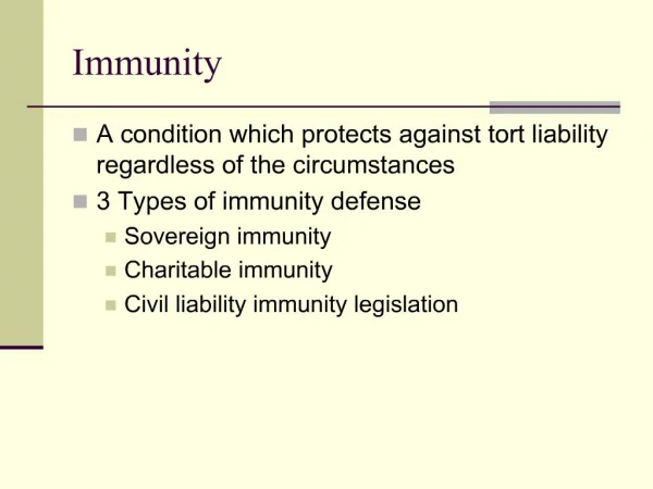
Transport Systems & Immunity. Chapters 42 & 43 Evolution & Types Main structures (Heart) Cardiac cycle Pathway of Blood flow Artery vs. Vein Blood Exchange @ Capillary level Lymphatic system Immune Response. Introductory Questions #1.

E N D
Transport Systems & Immunity • Chapters 42 & 43 • Evolution & Types • Main structures (Heart) • Cardiac cycle • Pathway of Blood flow • Artery vs. Vein • Blood • Exchange @ Capillary level • Lymphatic system • Immune Response
Introductory Questions #1 • Before any type of circulatory was established, how did organisms move substances throughout the body as with sponges, cnidarians, flatworms, and nematodes • Define the following: Hemolymph, hemocoel, hemocyanin, and interstitial fluid. • What is the difference between an open and closed circulatory system? • List all of the structures that a red blood cells will encounter as it circulates throughout the body beginning with the Vena cava. • Give three differences between an artery and a vein.
Circulation System Evolution • Simple diffusion: substances move from environment directly into the cells. (2 –3 cells thick) • Gastrovascular cavity (cnidarians, flatworms) • Open circulatory • hemolymph (blood & interstitial fluid) • sinuses (spaces surrounding organs): hemocoel • Closed circulatory: blood confined to vessels • Cardiovascular system • heart (atria/ventricles) • blood vessels (arteries, arterioles, capillary beds, venules, veins) • blood (circulatory fluid)
Several Types of Internal Transport have evolved in animals • In cnidarians and flatworms, the gastrovascular cavity functions in both • digestion • internal transport Mouth Circularcanal
Circulation System Evolution • Fish: • 2-chambered heart • single circuit of blood flow • Amphibians: • 3-chambered heart • 2 circuits of blood flow- • Circulation is “Pulmocutaneous” (lungs and skin) • Some mixing of blood • Mammals: • 4-chambered heart • Double circulation • Complete separation between oxygen-rich and oxygen poor blood
Open systems • A heart pumps blood through open-ended vessels into spaces between cells • Most animals have a separate circulatory system, either open or closed Tubular heart Pores Figure 23.2B
A heart pumps blood through arteries and capillary beds • The blood returns to the heart via veins • Closed systems Capillary beds Arteriole Artery(O2-rich blood) Venule Vein Atrium Heart Artery(O2-poor blood) Ventricle Gillcapillaries Figure 23.2C
Vertebrate cardiovascular systems reflect evolution Gill capillaries • A fish has a single circuit of blood flow Heart: Ventricle (V) Atrium (A) Systemic capillaries Figure 23.3A
Double circulation • From right ventricle to lungs via pulmonary arteries through semilunar valve (pulmonary circulation) • Capillary beds in lungs to left atrium via pulmonary veins • Left atrium to left ventricle (through atrioventricular valve) to aorta • Aorta to coronary arteries; then systemic circulation • Back to heart via two venae cavae (superior and inferior); right atrium
Pulmonaryartery Aorta Pulmonaryartery Superiorvena cava LEFTATRIUM RIGHTATRIUM Pulmonaryveins Pulmonaryveins Semilunarvalve Semilunarvalve Atrioventricularvalve Atrioventricularvalve Inferiorvena cava RIGHTVENTRICLE LEFTVENTRICLE Figure 23.4A
What is a heart attack? • A heart attack is damage that occurs when a coronary feeding the heart is blocked Aorta Rightcoronaryartery Leftcoronaryartery Blockage Dead muscle tissue Figure 23.8A
IQ #2 Pulmonaryartery 11. vessel Aorta 10. vessel Pulmonaryartery 1. vessel Superiorvena cava LEFTATRIUM 2. chamber RIGHTATRIUM Pulmonaryveins Pulmonaryveins 9. vessels 3. valve Semilunarvalve Semilunarvalve 8. valve Atrioventricularvalve 7. valve Atrioventricularvalve 4. vessel Inferiorvena cava 5. chamber RIGHTVENTRICLE 6. chamber LEFTVENTRICLE Figure 23.4A
Superiorvena cava 7 Capillaries of Head and arms Pulmonaryartery Pulmonaryartery Capillariesof right lung Capillariesof left lung Aorta 9 6 2 3 3 4 11 Pulmonaryvein Pulmonaryvein 5 LEFT ATRIUM 1 RIGHT ATRIUM LEFT VENTRICLE RIGHT VENTRICLE 10 Aorta Inferiorvena cava Capillaries ofabdominal organsand legs 8 Figure 23.4B
RBC Pathway through the Circulatory System Blood from Systemic Circuit Vena cava (inferior & superior) Right atrium (Tricuspid valve-AV valve) Right ventricle (Pulmonary semilunar valve) Pulmonary circuit –Lungs (P. arteries LungsP. veins) Left atrium (Bicuspid “Mitral” valve) Left Ventricle (Aortic semilunar valve) Aorta (arch, coronary, carotid, & abdominal, renal, mesenteric, iliac arteries)
Facts about the Circulatory System • Blood volume in the heart per contraction 70 ml (Stroke volume) • Total blood volume in a human 5 Liters (1.32 Gal) • Normal Beats per minute (BPM) 72 Bpm • Normal Blood pressure 120/80 mm Hg • Starling’s Law: when more blood is delivered to the heart, the heart stretches more and contracts with greater force which pumps more blood into arteries.
Cardiac Output • The volume of blood pumped out by the left ventricle • Determined by: (Stroke volume) x (Heart rate) Ex. 70 ml (per beat) x 72 BPM = 5040 ml/min Approx. 5 Liters per minute
The Heart Contracts and Relaxes Rhythmically 1 Heart isrelaxed.AV valvesare open. 2 Atriacontract. • Diastole • Blood flows from the veins into the heart chambers • Systole • The atria briefly contract and fill the ventricles with blood • Then the ventricles contract and propel blood out SYSTOLE 0.1 sec 3 Ventriclescontract.Semilunarvalvesare open. 0.3 sec 0.4 sec DIASTOLE Figure 23.6
The Heartbeat • Sinoatrial (SA) node (“pacemaker”): sets rate and timing of cardiac contraction by generating electrical signals • Atrioventricular (AV) node: relay point (0.1 second delay) spreading impulse to walls of ventricles • Electrocardiogram (ECG or EKG)
The Structure of blood vessels fits their Functions • A single layer of epithelial cells forms capillary walls • Arteries and veins have smooth muscle and connective tissue • Valves in veins prevent the backflow of blood
Blood Vessel Structural Differences • Capillaries •endothelium; basement membrane • Arteries •thick connective tissue; thick smooth muscle; endothelium; basement membrane • Veins•thin connective tissue; thin smooth muscle; endothelium; basement membrane
Cardiovascular disease • Cardiovascular disease (>50% of all deaths) • Heart attack- death of cardiac tissue due to coronary blockage • Stroke- death of nervous tissue in brain due to arterial blockage • Atherosclerosis: arterial plaques deposits • Arteriosclerosis: plaque hardening by calcium deposits • Hypertension: high blood pressure • Hypercholesterolemia: LDL, HDL
The circulatory system associates Intimately with all body tissues • Capillaries are microscopic blood vessels • They form an intricate network among the tissue cells Capillary Redbloodcell Figure 23.1A
Introductory Questions #1 • Before any type of circulatory was established, how did organisms move substances throughout the body as with sponges, cnidarians, flatworms, and nematodes • Define the following: Hemolymph, hemocoel, hemocyanin, and interstitial fluid. • What is the difference between an open and closed circulatory system? • List all of the structures that a red blood cells will encounter as it circulates throughout the body beginning with the Vena cava. • Give three differences between an artery and a vein.
Blood Exerts Pressure on Vessel Walls • Blood pressure depends on • Cardiac output • Blood volume • Resistance of vessels
Systolicpressure Diastolicpressure • Pressure is highest in the arteries • It drops to zero by the time the blood reaches the veins Relative sizes andnumbersof blood vessels Figure 23.9A
Connection: Measuring Blood Pressure can Reveal Cardiovascular Problems • Blood pressure is measured as systolic and diastolic pressures Blood pressure120 systolic80 diastolic(to be measured) Pressurein cuffabove120 Pressurein cuffbelow120 Pressurein cuffbelow 80 Rubber cuffinflated with air Soundsaudible instethoscope Soundsstop Arteryclosed Artery 2 3 4 1 Figure 23.10
Hypertension is persistent systolic pressure higher than 140 mm Hg and/or diastolic pressure higher than 90 mm Hg • It is a serious cardiovascular problem
muscle contractions • breathing • one-way valves • Three factors keep blood moving back to the heart Direction ofblood flowin vein Valve (closed) Valve (open) Skeletal muscle Figure 23.9B
Smooth Muscle Controls the Distribution of Blood • Muscular constriction of arterioles and precapillary sphincters controls the flow through capillaries Precapillary sphincters Thoroughfarechannel Thoroughfarechannel Venule Arteriole Venule Arteriole Capillaries Sphincters contracted 2 1 Sphincters relaxed
Introductory Questions #3 (See chapters 42 & 43) 1) In the cardiac cycle how is systole different from diastole? 2) Where is the SA and AV node located? What do these structures do? • Name three factors that can affect your blood pressure. • Blood is composed of a variety of things. Make a list of cellular and non-cellular substances present in blood. • Briefly explain how blood clots. (pg. 882) What proteins and cell parts are required for blood to clot? • What forces are involved in the exchange of gases and solutes at the capillary level? (pg. 879) • What areas of the body do we find a high number of lymph nodes?(pg. 901) • How are B cells different from T cells?
Review of Blood Pressure • Key factors that effect BP (CO, BV, R) • Regulated by a hormone called Renin • Renin released by the kidneys • Causes other proteins to increase in concentration and constrics the vessels -Angiotension -Aldosterone (hormone released by adrenal glands)
Pg. 880 Withdrawblood Centrifuge Place in tube PLASMA 55% CONSTITUENT MAJOR FUNCTIONS CELLULAR ELEMENTS 45% Solvent forcarrying othersubstances CELL TYPE NUMBER(per mm3 of blood) FUNCTIONS Water Erythrocytes(red blood cells) Salts 5–6 million Transport ofoxygen (and carbon dioxide) Sodium Potassium Calcium Magnesium Chloride Bicarbonate Osmotic balance,pH buffering, andregulation ofmembranepermeability Leukocytes(white blood cells) Defense andimmunity 5,000–10,000 Plasma proteins Albumin Fibrinogen Immunoglobins(antibodies) Osmotic balance,pH buffering Clotting Immunity Lymphocyte Basophil Eosinophil Substances transported by blood Monocyte Nutrients (e.g., glucose, fatty acids, vitamins) Waste products of metabolism Respiratory gases (O2 and CO2) Hormones Neutrophil Platelets 250,000–400,000 Blood clotting Figure 23.13
Red blood cells transport oxygen -Hemoglobin transport of O2 -Red blood cells contain hemoglobin (250-300 million) -RBC count: 4.2 – 6.2 million cells per mm3. (adult males & females) -Average Lifespan: 120 days -33% of RBC volume is hemoglobin -2.4 million are destroyed per second and are replaced in the bone marrow -No nucleus or mitochondria Figure 23.14
White blood cells help defend the body • White blood cells function both inside and outside the circulatory system • They fight infections and cancer Basophil Eosinophil Monocyte Lymphocyte Neutrophil Figure 23.15
WBC Type and Function • WBC count: 7000 per µL (1:700 RBC’s) • Neutrophils: most abundant phagocytic cells in the blood (60-70% of all WBC’s) • Eosinophils: containd oxidases & peroxidases -increase during allergic reactions -parasitic infections • Basophils: also important in allergic reactions -do not contain lysosomes -histamine in the cytoplasm (inflamm.) -heparin acts as an anticoagulant (prevents blood clots) • Lymphocytes: produce antibodies attack bacteria & viruses two types of cells form (B cells & T cells) • Monocytes: Largest of all WBC’s that become macrophages (about 5% of all WBC’s)
Stem cells offer a potential cure for leukemia and other blood cell diseases • All blood cells develop from stem cells in bone marrow • Such cells may prove valuable for treating certain blood disorders Figure 23.17
Blood clots plug leaks when blood vessels are injured • When a blood vessel is damaged, platelets respond • They help trigger the formation of an insoluble fibrin clot that plugs the leak Figure 23.16B
1 Injury to lining of bloodvessel exposes connectivetissue; platelets adhere 2 Platelet plug forms 3 Fibrin clot trapsblood cells Connectivetissue Plateletplug Platelet releases chemicalsthat make nearby platelets sticky Clotting factors from: Platelets Calcium andother factorsin blood plasma Damaged cells Pg. 882 Ca ions, clotting factors Prothrombin (Liver, Vit K) Thrombin Fibrinogen Fibrin Figure 23.16A
No substance has to diffuse far to enter or leave a cell Capillary Diffusion ofmolecules INTERSTITIALFLUID Tissuecell Figure 23.1B
leakage through clefts in the capillary walls • diffusion through the wall • blood pressure • osmotic pressure • The transfer of materials between the blood and interstitial fluid can occur by
Capillaries allow the Transfer of Substances Through Their Walls Figure 23.12A