Download
1 / 58
610 likes | 918 Views
Immunity. Innate immunity : defenses against any pathogen Adaptive immunity : induced resistance to a specific pathogen. ANIMATION Host Defenses: The Big Picture. Historical Development. Pasteur observed immunity in chickens injected with weakened pathogens

E N D
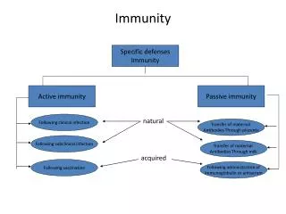
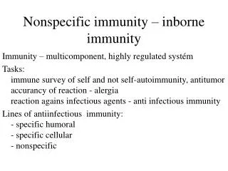
Immunity • Innate immunity: defenses against any pathogen • Adaptive immunity: induced resistance to a specific pathogen ANIMATION Host Defenses: The Big Picture
Historical Development • Pasteur observed immunity in chickens injected with weakened pathogens • Von Behring received the Nobel prize for development of antitoxin • Ehrlich’s work led to the identification of antibodies in serum
Figure 17.20 The dual nature of the adaptive immune system. Humoral (antibody-mediated) immune system Cellular (cell-mediated) immune system Control of freely circulating pathogens Control of intracellular pathogens Intracellular antigens are expressed on the surface of an APC, a cell infected by a virus, a bacterium, or a parasite. Extracellular antigens A B cell binds to the antigen for which it is specific. A T-dependent B cell requires cooperation with a T helper (TH) cell. A T cell binds to MHC–antigen complexes on the surface of the infected cell, activating the T cell (with its cytokine receptors). T cell Cytokines activate T helper (TH) cell. Cytokines activate macrophage. Cytokines Cytokines B cell Cytokines from the TH cell transform B cells into antibody-producing plasma cells. Activation of macrophage (enhanced phagocytic activity). The B cell, often with stimulation by cytokines from a TH cell, differentiates into a plasma cell. Some B cells become memory cells. TH cell The CD8+T cell becomes a cytotoxic T lymphocyte (CTL) able to induce apoptosis of the target cell. Cytotoxic T lymphocyte Plasma cell Plasma cells proliferate and produce antibodies against the antigen. Memory cell Some T and B cells differentiate into memory cells that respond rapidly to any secondary encounter with an antigen. Lysed target cell
Dual Nature of Adaptive Immunity • T and B cells develop from stem cells in red bone marrow
Figure 17.8 Differentiation of T cells and B cells. Stem cells develop in bone marrow or in fetal liver Stem cell (diverges into two cell lines) Red bone marrow of adults Thymus Differentiate to B cells in adult red bone marrow Differentiate to T cells in thymus B cell T cell Migrate to lymphoid tissue such as spleen, but especially lymph nodes
Dual Nature of Adaptive Immunity • Humoral immunity • Due to antibodies • B cells mature in the bone marrow • Chickens: bursa of Fabricius ANIMATION Humoral Immunity: Overview
Dual Nature of Adaptive Immunity • Cellular immunity • Due to T cells • T cells mature in the thymus
The Nature of Antigens • Antigen (Ag): a substance that causes the body to produce specific antibodies or sensitized T cells • Antibodies (Ab) interact with epitopes, or antigenic determinants • Hapten: antigen is combined with carrier molecules
Figure 17.1 Epitopes (antigenic determinants). Antibody A Epitopes (antigenic determinants) on antigen Antigens:componentsof cell wall Binding sites Bacterial cell Antibody B
Figure 17.2 Haptens. Hapten-carrier conjugate Hapten molecules Carrier molecule
The Nature of Antibodies • Globular proteins called immunoglobulins • The number of antigen-binding sites determines valence
Figure 17.3ab The structure of a typical antibody molecule. Antigen-binding site Heavy chain Light chain Fc (stem) region Hinge region Antibody molecule Epitope (antigenic determinant) Antigen Antigen- binding site Enlarged antigen-binding site bound to an epitope
Figure 17.3c The structure of a typical antibody molecule. Antibodies Antibody molecules shown by atomic force microscopy (see page 64)
IgG Antibodies • Monomer • 80% of serum antibodies • Fix complement • In blood, lymph, and intestine • Cross placenta • Enhance phagocytosis; neutralize toxins and viruses; protect fetus and newborn • Half-life = 23 days
IgM Antibodies • Pentamer • 5–10% of serum antibodies • Fix complement • In blood, in lymph, and on B cells • Agglutinate microbes; first Ab produced in response to infection • Half-life = 5 days
Chapter 17, unnumbered figure, page 483. Disulfide bond J chain
IgA Antibodies • Dimer • 10–15% of serum antibodies • In secretions • Mucosal protection • Half-life = 6 days
Chapter 17, unnumbered figure, page 483. J chain Secretory component
IgD Antibodies • Monomer • 0.2% of serum antibodies • In blood, in lymph, and on B cells • On B cells, initiate immune response • Half-life = 3 days
IgE Antibodies • Monomer • 0.002% of serum antibodies • On mast cells, on basophils, and in blood • Allergic reactions; lysis of parasitic worms • Half-life = 2 days
Activation of B Cells • Major histocompatibility complex (MHC) expressedon mammalian cells • T-dependent antigens • Ag presented with (self) MHC to TH cell • TH cell produces cytokines that activate the B cell • T-independent antigens • Stimulate the B cell to make Abs ANIMATION Antigen Processing and Presentation: Overview
Figure 17.6 T-independent antigens. Polysaccharide (T-independent antigen) Epitopes B cell receptors
Figure 17.4 Activation of B cells to produce antibodies. Extracellularantigens MHC class II with Ag fragment displayed on surface MHC class II with Ag fragment Antibodies Ag fragment B cell B cell Immunoglobulin receptorscoatingB cell surface Plasma cell TH cell B cell Cytokines Immunoglobulin receptors on B cell surface recognize and attach to antigen, which is then internalized and processed. Within the B cell a fragment of the antigen combines with MHC class II. MHC class II–antigen-fragment complex is displayed on B cell surface. Receptor on the T helper cell (TH) recognizes complex of MHC class II and antigen fragment and is activated— producing cytokines, which activate the B cell. The TH cell has been previously activated by an antigen displayed on a dendritic cell (see Figure 17.10). B cell is activated by cytokines and begins clonal expansion. Some of the progeny become antibody-producing plasma cells.
Figure 17.5 Clonal selection and differentiation of B cells. Stem cell Stem cells differentiate into mature B cells, each bearing surface immunoglobulins against a specific antigen. Antigen B cell III complexes with its specific antigen and proliferates. B cells II I III IV Memory cells Some B cells proliferate into long-lived memory cells, which at a later date can be stimulated to become antibody-producing plasma cells. See Figure 17.17. Other B cells proliferate into antibody-producing plasma cells. Plasma cells Plasma cells secrete antibodies into circulation. Antigens in circulation now attached to circulating antibodies Cardiovascular system
Activation of B Cells • B cells differentiate into: • Antibody-producing plasma cells • Memory cells • Clonal deletion eliminates harmful B cells
Antigen–Antibody Binding • Agglutination • Opsonization • Activation of complement • Antibody-dependent cell-mediated cytotoxicity • Neutralization ANIMATION Humoral Immunity: Antibody Function
Figure 17.7 The results of antigen–antibody binding. PROCTECTIVE MECHANISM OF BINDING ANTIBODIES TO ANTIGENS Agglutination (see also Figure 18.5) Activation of complement (see also Figure 16.9) Causes inflammation and cell lysis Reduces number of infectious units to be dealt with Complement Bacteria Lysis Bacterium Antibody-dependent cell-mediated cytotoxicity (see also Figure 17.16) Opsonization (see also Figure 16.9) Coating antigen with antibody enhances phagocytosis Antibodies attached to target cell cause destruction by macrophages, eosinophils, and NK cells Phagocyte Eosinophil Epitopes Large target cell (parasite) Neutralization (see also Figure 18.9) Perforin and lytic enzymes Blocks adhesion of bacteria and viruses to mucosa Blocks attachment of toxin Virus Toxin Bacterium
T Cells and Cellular Immunity • T cells mature in the thymus • Thymic selection eliminates many immature T cells
Table 17.2 Principal Cells That Function in Cell-Mediated Immunity
T Cells and Cellular Immunity • T cells respond to Ag by T-cell receptors (TCRs) • T cells require antigen-presenting cells (APCs) • Pathogens entering the gastrointestinal or respiratory tracts pass through: • M (microfold) cells over • Peyer’s patches, which contain APCs
(a) M cell on Peyer’s patch. Note the tips of the closely packed microvilli on the surrounding epithelial cells. (b) M cells facilitate contact between the antigens passing through the intestinal tract and cells of the body’s immune system. Figure 17.9 M cells. Microvilli on epithelial cell Antigen M cell TH cell Pocket B cells Macrophage Epithelial cell
T Helper Cells • CD4+ or TH cells • TCRs recognize Ags and MHC II on APC • TLRs are a costimulatory signal on APC and TH • TH cells produce cytokines and differentiate into: • TH1cells • TH2 cells • TH17 cells • Memory cells
TH1 produce IFN-gwhich activates cells related to cell-mediated immunity, macrophages, and Abs TH2 activate eosinophils and B cells to produce IgE TH17 stimulate the innate immune system TF stimulate B cells to produce plasma cells and are involved in class switching T Helper Cells ANIMATION Antigen Processing and Presentation: Steps
Figure 17.11 Lineage of effector T helper cell classes and pathogens targeted. Antibodies TH1 cells B cell TH2 cells TH17 cells Cell-mediated immunity; control of intracellular pathogens, delayed hypersensitivity reactions (page 535); stimulates macrophages. Recruits neutrophils; provides protection against extracellular bacteria and fungi TH cell IL-17 IFN-g TH17 cells TH1 cells IL-4 TH2 cells Fungi Extracellular bacteria Neutrophil Macrophage Intracellular bacteria and protozoa Mast cell Basophil Eosinophil Important in allergic responses, especially by production of IgE Stimulates activity of eosinophils to control extracellular parasites such as helminths (see ADCC, page 495). Helminth
Figure 17.10 Activation of CD4+T helper cells. An APC encounters and ingests a microorganism. The antigen is enzymatically processed into short peptides, which combine with MHC class II molecules and are displayed on the surface of the APC. A receptor (TCR) on the surface of the CD4+T helper cell (TH cell) binds to the MHC–antigen complex. If this includes a Toll-like receptor, the APC is stimulated to secrete a costimulatory molecule. These two signals activate the TH cell, which produces cytokines. TH cell receptor (TCR) The cytokines cause the TH cell (which recognizes a dendritic cell that is producing costimulatory molecules) to become activated. APC (dendritic cell) T helper cell Antigen Complex of MHC class II molecule and antigen fragment Cytokines Antigen fragment (short peptides) Microorganism carrying antigens Costimulatory molecule, (required to activate T cells that have not previously encountered antigen)
T Cytotoxic Cells • CD8+or TC cells • Target cells are self-cells carrying endogenous antigens • Activated into cytotoxic T lymphocytes (CTLs) • CTLs recognize Ag + MHC I • Induce apoptosis in target cell • CTL releases perforin and granzymes ANIMATION Cell-Mediated Immunity: Cytotoxic T Cells
Figure 17.12 Killing of virus-infected target cell by cytotoxic T lymphocyte. Processed antigen presented with MHC class I T cell receptors Infected target cell is lysed MHC class I Processed antigen CTL Virus-infected cell (example of endogenous antigen) Virus-infected cell Cytotoxic T lymphocyte (CTL) A normal cell will not trigger a response by a cytotoxic T lymphocyte (CTL), but a virus-infected cell (shown here) or a cancer cell produces abnormal endogenous antigens. The abnormal antigen is presented on the cell surface in association with MHC class I molecules. CD8+T cells with receptors for the antigen are transformed into CTLs. The CTL induces destruction of the virus-infected cell by apoptosis.
T Regulatory Cells • Treg cells • CD4 and CD25 on surface • Suppress T cells against self
Antigen-Presenting Cells • Digest antigen • Ag fragments on APC surface with MHC • B cells • Dendritic cells • Activated macrophages ANIMATION Antigen Processing and Presentation: MHC
Figure 17.15 Activated macrophages. Activated macrophages Resting (inactive) macrophage
Natural Killer (NK) Cells • Granular leukocytes destroy cells that don’t express MHC I • Kill virus-infected and tumor cells • Attack parasites
ADCC • Antibody-dependent cell-mediated cytotoxicity
Figure 17.16 Antibody-dependent cell-mediated cytotoxicity (ADCC). KEY Macrophage Cytotoxic cytokines Lytic enzymes Perforin enzymes Eosinophil Extracellular damage Fc region Large parasite Epitope Antibody (a) Organisms, such as many parasites, that are too large for ingestion by phagocytic cells must be attacked externally. Eosinophils Fluke (b) Eosinophils adhering to the larval stage of a parasitic fluke
Figure 17.16a Antibody-dependent cell-mediated cytotoxicity (ADCC). KEY Macrophage Cytotoxic cytokines Lytic enzymes Perforin enzymes Eosinophil Extracellular damage Fc region Epitope Large parasite Antibody (a) Organisms, such as many parasites, that are too large for ingestion by phagocytic cells must be attacked externally.
Figure 17.16b Antibody-dependent cell-mediated cytotoxicity (ADCC). Eosinophils Fluke Eosinophils adhering to the larval stage of a parasitic fluke.