Download
1 / 2
30 likes | 490 Views
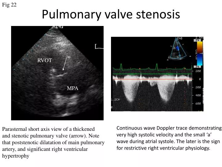
Fig 22. Pulmonary valve stenosis. RVOT. MPA. Continuous wave Doppler trace demonstrating very high systolic velocity and the small ‘ a ’ wave during atrial systole. The later is the sign for restrictive right ventricular physiology. Parasternal short axis view of a thickened

E N D
Fig 22 Pulmonary valve stenosis RVOT MPA Continuous wave Doppler trace demonstrating very high systolic velocity and the small ‘a’ wave during atrial systole. The later is the sign for restrictive right ventricular physiology. Parasternal short axis view of a thickened and stenotic pulmonary valve (arrow). Note that poststenotic dilatation of main pulmonary artery, and significant right ventricular hypertrophy
Fig 23 Pulmonary Regurgitation A B Continuous wave Doppler recording obtained from the parasternal position directed through pulmonary valve. A: From a patient with mild PR, note the regurgitation signal persist till the end of diastole. B: Patient with severe pulmonary regurgitation; the regurgitation signal ends at the middle of diastole.