Download
1 / 37
370 likes | 687 Views
Physician: Mark D. Plunkett, M.D. Author: Heather Nolan, BA, AS. Pulmonary valve REPLACEMENT WITH BENTALL/AORTIC VALVE AND ROOT REPLACEMENT. UK. Center for Minimally Invasive Surgery. Patient Presentation. For Aortic Root Repair/Replacement Aortic root aneurysm

E N D
Physician: Mark D. Plunkett, M.D. Author: Heather Nolan, BA, AS Pulmonary valve REPLACEMENT WITH BENTALL/AORTIC VALVE AND ROOT REPLACEMENT UK Center for Minimally Invasive Surgery
Patient Presentation For Aortic Root Repair/Replacement • Aortic root aneurysm • Aortic dissection affecting both root and valve • Symptoms: cough, diastolic murmur, dysphasia, dyspnea on exertion, fatigue, palpitations, and widened pulse pressures For Aortic Valve Repair/Replacement • Aortic valve stenosis • Aortic valve insufficiency • Aortic regurgitation • Symptoms: angina, dizziness, fainting, fatigue, shortness of breath, swelling of ankles and legs, arrhythmia, and palpitations For Pulmonary Valve Repair/Replacement • Pulmonary valve stenosis • Pulmonary valve insufficiency • Pulmonic regurgitation • Symptoms: dyspnea, angina, cyanosis, congestive heart failure, fatigue, fluid retention, cough, cardiomegaly, and syncope
Patient work-up • Chest X-Ray • Electrocardiogram • Echocardiogram • Cardiac Catheterization
OR set-up: Personel Echo Techs Surgeon 1 Anesthesiologist Surgeon 2 Scrub Tech 2 Perfusionist Scrub Tech 1
OR set-up: surgical Equipment Anesthesia Workstation Transesophageal Echocardiograph Heart-lung Machine Continue to Video Equipment
Anesthesia workstation • For more information: www.udmercy.edu/crna/agm/02.htm Back to Surgical Equipment
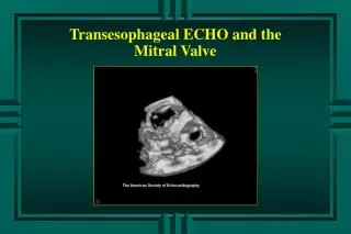
Transesophageal echocardiograph • For more information: www.med.yale.edu/intmed/cardio/imaging/techniques/echo_tee/index.html Back to Surgical Equipment
Heart-lung machine • For more information: www.surgeryencyclopedia.com/Fi-La/Heart-Lung-Machines.html Back to Surgical Equipment
OR set-up: video Equipment Camera Monitors 45° Laparascope (optional)
Or set-up: instruments Click on + for individual instruments • Kellys, Tonsils, Kockers, Allis • Clamps + • Scissors + • Sponge sticks • Tubing clamps • Wire cutter • Malleables + • Weitlanders • Suction tips + • Suction tube • Tube holder • Cross-clamp • Sternal wire (needle included) • Hemoclip appliers + • Vascular clamps + • Forceps + • Needle holders + • Handles + • Nerve hooks + • Penfield • Tourniquets • Dilators + • Spring instruments + • Retractors + • Defibrillator paddles • Mosquitos + • Hemostats Continue to Procedure Steps
instruments: Hemoclip appliers • Small hemoclip appliers • Medium hemoclip appliers • Short yellow hemoclip appliers Back to Instruments
instruments: vascular clamps • Left blue titanium vascular clamp • Right blue titanium vascular clamp • Deborah Castaneda clamp • Castaneda anastomosis clamp • Derra partial occlusion clamp • Cooley derra clamp • Debakey anastamosis clamp • Debakey general purpose clamp • Straight Debakey bulldog clamp • Debakey spoon perivascular clamp • Debakey multipurpose clamp • Debakey acutely curved clamp • Angled Debakey Back to Instruments
instruments: forceps • Adson tissue forceps with teeth • Gerald forceps • Debakey forceps • Scanlon smooth tip forceps • Scanlon Debakey fine forceps Back to Instruments
instruments: needle holders • Castro round handle locking needle holder • Non-locking castro round handle needle holder • Fine tip Scanlon needle holder • Sarot needle holder • Creelewood needle holder • Small Berry needle holder Back to Instruments
instruments: handles • Beaver handle • Knife handle Back to Instruments
instruments: nerve hooks • Dandy nerve hook • Dull nerve hook • Sharp nerve hook • Mid tip Janetta right angle hook Back to Instruments
instruments: dilators • 1.0 dilator • 1.5 dilator • 2.0 dilator • 2.5 dilator • 3.0 dilator • Joseph hook single prong • Aortic arch dilator Back to Instruments
instruments: spring instruments • Spring Potts scissor flat handle • Spring Potts scissor round handle • Small Dietrich bulldog Back to Instruments
instruments: retractors • Kirkland retractor • ALM retractor • Finochetto retractor • Morse retractor • Chest retractor • Ragnell retractor • Sharp Senn retractor • Short sharp rake • Vein retractor • Army-Navy retractor • Ankenney retractor • Touffier retractor • Dr. Salley retractor Back to Instruments
instruments: mosquitos • CVD mosquito • STR mosquito • Jacobson mosquito Back to Instruments
instruments: clamps • Jacobs clamp • Pennington clamp • Right angle clamp • Small tubing clamp • Medium tubing clamp • Peer towel clamps Back to Instruments
instruments: scissors • STR Mayo scissors • CVD Mayo scissors • Metz scissors • Curved fine Cooley scissors • Curved heavy Cooley scissors • Demartel scissors • Jamison black handle supercut scissors • Straight Mayo Harrington scissors • Wire scissors • Pump line scissors Back to Instruments
instruments: malleables • 1/4” malleable • 5/8” malleable • 3/8” malleable • 1/2” malleable • 3/4” malleable • 1” malleable Back to Instruments
instruments: suction tips • Boss pump suction tip • Frazier suction tip • Yankauer suction tip Back to Instruments
Make sternotomy incision (more) Place heart on cardiopulmonary bypass (more) Add cardioplegic agent (more) Expose and remove aortic valve Size aortic replacement valve (more) Expose and remove pulmonic valve Size pulmonic valve replacement (more) Attach aortic valve and root replacement to heart (more) Expose left coronary artery Expose and trim native aortic root Attach pulmonary homograft (more) Attach coronary arteries to aortic root replacement (more) Attach aortic root replacement to ascending aorta (more) Take heart off cardiopulmonary bypass (more) Close (more) Procedure Steps UK Continue to Post-Operative Care Center for Minimally Invasive Surgery
Sternotomy incision • Make incision along sternal midline using scalpel • Cauterize any bleeding vessels • Use sternal saw to cut sternum • Apply bone paste to cut edge of sternum • Place sterile towels on cut edge of sternum • Use retractors to access surgical area Back to Procedure Steps
Cardiopulmonary bypass (starting) • Put purse string suture into superior vena cava • Thread suture through tourniquet and secure with hemostat • Cut vein wall • Insert bypass cannula into vein • Cinch tourniquet and secure with hemostat • Repeat above to inferior vena cava • Repeat for aorta • Attach retrograde cardioplegia • Cross-clamp aorta • Connect cannulae to bypass tubing Back to Procedure Steps
Cardiopulmonary bypass (removing) • Uncinch tourniquet of superior vena cava • Remove cannula while simultaneously tightening purse string sutures • Add 6 knots to purse string suture • Repeat for inferior vena cava • Repeat for aorta Back to Procedure Steps
Cardioplegic agent For Antegrade Cardioplegia • Once aortic valve is removed, administer cardioplegic agent at the aortic root • Repeat approximately every 20-30 minutes For Retrograde Cardioplegia • use purse string sutures to place retrograde cannula in coronary sinus • Cardioplegic agent is administered continuously • Remove cannula, close purse strings, tie off suture Antegrade versus Retrograde Considerations • Size of anatomy • Length of procedure • Access to coronary sinus and aortic root Back to Procedure Steps
Sizing replacement valves • Replacement valve sizes range from 16-33 mm • These sizes are measured as the external diameter of the prosthetic valve with the sewing ring compressed • Use a valve sizing tool (pictured) to get optimal size • Match the replacement size to the native valve • Error on the large side to get the largest possible diameter for maximal blood flow • Check valve function prior to placement Back to Procedure Steps
Bentall aortic root replacement • Replacement includes valve and root • When removing native aorta/aortic root detach the coronary ostia (opening) from the aorta leaving a small rim of aortic tissue (Note: this is deemed the “button”) • Size aortic replacement device (valve and root combination) • Suture the device to the aortic annulus (fibrous tissue ring surrounding the opening to the aorta) (more) • Cut two holes in the root replacement for the coronary ostia using a thermal cutter or blade • Suture the coronary ostia to the root replacement (more) • Trim the root replacement to size • Suture root replacement to native ascending aorta Back to Procedure Steps
Suture: device-annulus • Use pledgetted sutures • Run end one of suture through the annulus starting from under the annulus • Run end one of suture through device’s sewing ring starting from under the device • Repeat with end two so that pledgett is up against underside of annulus • Alternate suture colors to allow for easier manipulation and tying of the device to the annulus • Push device into the annulus using previously placed suture to guide the device into place • Tie sutures in place (animation) Needle Pledgett Annulus Suture Back to Bentall Steps
Suture: device-ostia • Use a teflon strip for reinforcement • Place ostia within pre-cut opening in an end-to-side manner • Use a running stitch to secure ostia to device Back to Bentall Steps
Pulmonary homograft • Replacement valves can be biological or mechanical Biological Replacement Valves • A homograft, or allograft, comes from a human donor (pictured-in forceps) • A xenograft comes from animal tissue • Another option for aortic valve replacement is a pulmonary autograft in which the aortic valve is replaced with the patient’s native pulmonary valve Mechanical Replacement Valves • Are manmade and come in a variety of designs and materials Biological Versus Mechanical Considerations • Biologic valves reduce the risk of associated clots but are not as durable • Mechanical valves theoretically will last forever; however, there is an increased risk of clotting on the prostheses which can lead to stroke Back to Procedure Steps
Closing • Place external pacemaker leads • Check pacemaker leads and pacing • Place chest tube drainage cannulae • Prepare exposed sternal bone for closure using bone paste and electrocautery • Use sternal wires with the attached needle to close the sternum • Twist sternal wires together (twist number varies but 3-4 is recommended for optimal strength) • Bend exposed metal ends of sternal wire toward sternum • Close fascia • Close skin Back to Procedure Steps
Post-operative care • Connect patient to ventilator • Monitor ECG, oxygen saturation, blood pressures, and blood gases • Check urinary output and chest tube output • Prior to discharge: wean off ventilator, train patient on incentive spirometer, anticoagulation therapy, diet as tolerated, and ambulation
Possible complications • Thrombosis • Valve malfunction/failure • Root replacement malfunction/failure • Infection • Arrythmia • Death