Download
1 / 80
850 likes | 1.05k Views
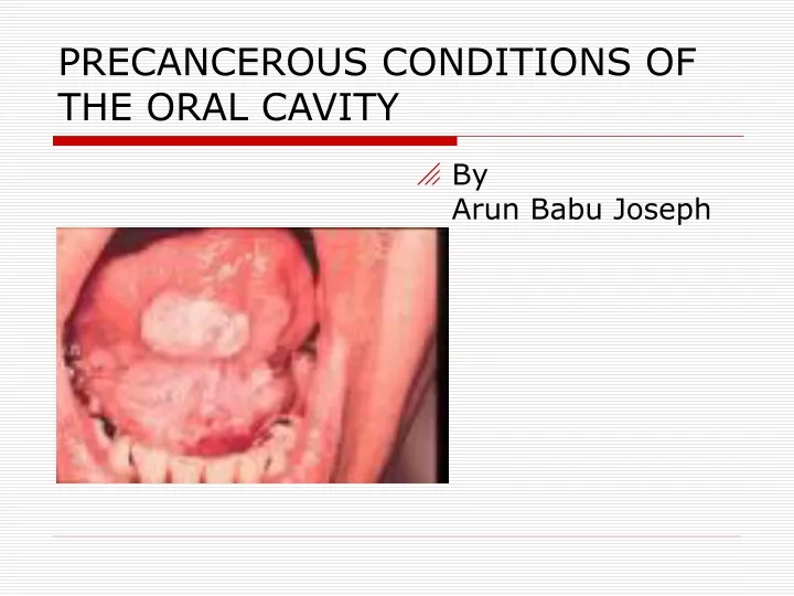
PRECANCEROUS CONDITIONS OF THE ORAL CAVITY. By Arun Babu Joseph. Classification ( Bailey). Lesions considered having a definite risk of malignancy Leukoplakia Erythroplakia Chronic hyperplastic candidiasis

E N D
PRECANCEROUS CONDITIONS OF THE ORAL CAVITY • By Arun Babu Joseph
Classification ( Bailey) Lesions considered having a definite risk of malignancy • Leukoplakia • Erythroplakia • Chronic hyperplastic candidiasis Conditions that are not themselves premalignant but which are associated with a higher than normal incidence of oral cancer • Oral submucous fibrosis • Syphilitic glossitis • Sideropenic dysphagia Oral conditions about which there is still some doubt as to whether their association with oral cancer is causal or casual • Oral Lichen planus • Discoid lupus erythematosus • Dyskeratosis congenita
Types of oral precancer. Precancerous lesions • Defined as morphologically altered tissue in which cancer is more likely to occur than its apparently normal counterpart. Example • Leukoplakia • Erythroplakia • Mucosal changes associated with smoking habits. • Carcinoma in situ • Bowen disease • Actinic keratosis, cheilitis and elastosis.
Precancerous conditions Defined as generalized state or condition associated with significantly increased risk for cancer development. Example Oral sub mucous fibrosis Syphilis Sideropenic dysphagia Oral Lichen planus Lupus erythematosus
Leukoplakia • Any white patch or plaque that cannot be characterized clinically or pathologically as any other disease. • This definition has no histological connotation.
Forms of leukoplakia.(WHO 1980) Homogenous : lesions that are uniformly white. Non homogenous : lesions in which part of the lesion is white and the rest appears reddened. Alternatively 1.) Homogenous a.) Smooth b.)Furrowed(fissured) c.) Ulcerated. 2.) Non homogenous nodulospeckled: Well demarcated raised white areas interspersed with reddened areas.
According to risk of future development of oral cancer. High risk sites • Floor of mouth • Lateral or ventral surface of tongue. • Soft palate. Low risk sites • Dorsum of tongue • Hard palate Intermediate group • All other sites of oral mucosa.
Etiology Local factors • Tobacco Smokeless and smoking tobacco. • Alcohol • Chronic irritation malocclusion ill fitting dentures, sharp broken teeth, hot spicy food, root piece etc • Candidiasis • Electromagnetic reaction or galvanism
Etiology (contd.) Regional & systemic factors. • Vitamin deficiency • Sideropenic anaemia • Nutritional deficiency • Conditions causing xerostomia ( Salivary gland diseases, Anticholinergic drugs, Radiation.) • Drugs ( Anticholinergics, Antimetabolic, Systemically administered alcohol) • Virus ( HSV & HPV) • Idiopathic
Histopathological features. Hallmark • Surface hyperkeratosis • Epithelial hyperplasia • Epithelial dysplasia
Keratinization pattern Keratinization of the mucosal epithelium which is normally non-keratinized. Hyperorthokeratosis • Orthokeratosis: In normal state , superficial epithelium, is nearly homogenous, eosinophilic and anuclearwith stratum granulosum always present. • Hyperorthokeratosis: abnormal increase in thickness of the orthokeratin layer of stratum corneum in a particular location. Hyperparakeratosis • Parakeratosis: superficial epithelium is flat and acidophilic with pyknotic nuclei. Stratum granulosum may or may not be present. • Hyperparakeratosis: Increased thickness of parakeratotic layer, exceeding the normal thickness.
Epithelium Epithelial thickness • Hyperplasia • Atrophy • Vacuolar degeneration • Acanthosis • Basal cell hyperplasia • Intra-epithelial edema
Epithelial dysplasia • Drop shaped rete ridges • Nuclear hyperchromatism • Nuclear pleomorphism • Altered nuclear-cytoplasmic ratio • Excess mitotic activity • Loss of polarity of cells • Deep cell keratinisation • Loss of differentiation • Loss of intercellular distance
Epithelial dysplasia seen in 3% of snuff induced leukoplakias and 16% of smoking habit related leukoplakia. • Nodular leukoplakia shows higher frequency of epithelial dysplasia • Grading
Connective tissue • Chronic inflammatory cell infiltration is seen in 50% cases. • Submucosal, homogenous eosinophilic material is usually seen in the connective tissue. • Hyaline degeneration seen in 10 % cases
Malignant potential • 0.3% - 10% cases • Higher in women (6%) than men (3.9%) due to involvement of endogenous factors • Leukoplakia associated with chewing habit of tobacco shows higher rate of malignant transformation. • Nodular dysplasia has higher risk of malignant transformation than other clinical types.
High risk if Elderly patient Persistence of lesion for several years Female patient Lesion situated on the margins, base of tongue, floor of mouth Erosive lesions
Differential diagnosis • Lichen planus • Chemical burns • Syphilitic mucus patches • White sponge nevus • Discoid lupus erythematosus • Psoriasis
Differential diagnosis (contd.) • Leukoedema • Hairy leukoplakia • Verruca vulgaris • Cheek biting lesion • Electrogalvanic white lesion
Management • Elimination of the etiological factor • Conservative treatment • 13 cis retinoic acid • Antioxidant therapy • Nystatin therapy
Surgical management Biopsy taken for microscopic examination from areas with greater surface irregularities. Conventional Surgery Cryosurgery Electrocautery LASER
Guidelines for treatment • Biopsy should be done • Elimination of etiological factors • Conservative /Surgical management if not heal in 2-3 weeks • Conservative treatment applied to large incipient lesions & verrucous lesions
Surgical treatment if conservative treatment fails in 3 months • Excision of nodular leukoplakia & follow up • All patients , re-examination twice a year • Re-biopsy after 5-6 months
ERYTHROPLAKIA • Also called erythroplasia of Queyrat. • Def: Area of reddened, velvety textured mucosa that cannot be identified on the basis of clinical & histopathologic examination as being caused by inflammation or any other disease process.
Etiology • Idiopathic • Alcohol • Smoking • Secondary infection or superinfection with candidiasis
Clinical features Seen as a non elevated red macule on an epithelial surface. Otherwise asymtomatic. • Age and sex Male predilection 6th & 7th decade • Sites • All mucosal surfaces of head & neck area • 50% found on vermilion or intraoral surfaces rest evenly divide between larynx & pharynx • Intraorally lateral & ventral tongue, oral floor & soft palate are more frequently involved.
Hisopathological features • Epithelial dysplasia • Cause for the red colour • Spinous layer contains cells showing atypia, hyperchromatism, pleomorphism, & increase in the number of mitotic figures.
Differential diagnosis. • Candidiasis • Denture stomatitis • Tuberculosis • Histoplasmosis • Area of mechanical irritation • Macular hemangioma • Telengiectasia. • Traumatic lesion
Management • 1 % toluidine blue test. • Incisional biopsy for microscopic diagnosis. • Conservative surgical procedure such as mucosal stripping • Destructive techniques such as electrocoagulation, cryotherapy, laser ablation also effective. • Extended clinical follow up. • Elimination of a suspect irritant
PIPE SMOKERS’ KERATOSIS • palatal keratosis due to pipe smoking is benign. • Any carcinomas related to pipe-smoking appears in another site in the mouth and may not be preceded by keratosis.
SMOKELESS TOBACCO-RELATED KERATOSES • Hyperkeratotic mucosal lesions Management • Diagnosis is based on the history of snuff use and the white lesion in the area where the tobacco is held. • Biopsy is required • Snuff-dippers’ lesions will resolve on stopping the habit • Regular follow-up
Bowens disease • Localized intra-epidermoid carcinoma • Characterized by progressive scaly or crusted plaque like lesion Causes • Sun exposure • Arsenic ingestion
Clinical features • Sites :male and female genital mucosa and in oral mucosa as erythroplakia, leukoplakia or eryhematous lesion. • Skin: red & slightly scaly area on the skin, which eventually enlarges and turns into white or yellowish lesion. • Signs: when scales are removed a granular surface without bleeding is seen.
Histopathology Intraepithelial features of malignancy Management Freezing technique/diathermy/ cauterization/ radiotherapy/ application of cytotoxic drugs.
Chronic hyperplasic candidiasis. • Dense chalky plaques of keratin • Plaques thicker & more opaque than leukoplakia • Commonly seen at the oral commisures, extending into adjacent skin & face. • High degree of malignant change
Treatment nystatin, amphotercin, or miconazole to eliminate the infection Treatment necessary for many months & reinfection common Surgical excision for persistent lesions.
Oral Lichen planus • First described by Erasmus Wilson in 1869 • Inflammatory condition of skin presenting with characteristic violaceous , polygonal, pruritic papules. Lace like pattern. • Buccal mucosa ( 84%) lips, tongue, gingiva, floor of mouth & palate.
Etiology • Immunology Cell mediated immune response Autoimmunity Immunodeficiency • Genetic factors • Infections • Drugs & chemicals
Psychogenic factors Habit Miscellaneous Deficiency of vitamin B1, B6, & C,electric potential difference, anaemia, & patients with secondary syphilis. Trauma & malnutrition
Clinical features • Burning pain of the oral mucosa Appearance • Oral lesion characterized by white & gray velvety thread like papules in a linear angular or retiform arrangement forming typical lacy reticular patterns, rings and streaks over the oral mucosa. • Wickham’s striae--- tiny white elevated dots present at the intersection of white lines.
Types of oral lichen planus • Reticular type • Papular • Plaque • Atrophic form • Bullous form • Hypertrophic form • Annular form
Histopathological features of oral lichen planus • Hyperkeratosis • Prominent granular layer • Basal cell degeneration (may form colloid bodies) • Heavy lymphocyte infiltration (T cells) in the upper epidermis • Saw tooth dermo-epidermal junction.
Differential diagnosis • Leukoplakia • Candidiasis • Pemphigus • Lupus erythematosus • Drug induced lesion • White sponge nevus • Ectopic geographic tongue • Cheek biting • Lichenoid drug reaction
Malignant potential • 1.2 % malignant change (Silverman et al) • Association between LP & oral cancer seen only with atrophic or erosive LP.
Management • Removal of cause • Medical therapy Steroids In most patients with erosive & ulcerative lesion topical steroids are commonly used. If severe, systemic steroids are used. Topical Antifungal agents. Vitamin A analogues Cyclosporine
Surgical therapy. Indications When the conventional methods fail in ulcerative lesions In case of small solitary lesions Cryosurgery & cauterization have also been tried.