Download

1 / 18
250 likes | 1.56k Views
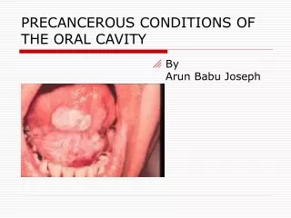
PREMALIGNANT CONDITIONS OF ORAL CAVITY. BY DR. MANISHA MISHRA. Types:. Oral submucous fibrosis Leukoplakia Erythroplakia Candidiasis. Leukoplakia. The term leukoplakia describes a greyish white patch or plaque found in the mucous membrane of the oral cavity.

E N D
PREMALIGNANT CONDITIONS OF ORAL CAVITY BY DR. MANISHA MISHRA
Types: • Oral submucous fibrosis • Leukoplakia • Erythroplakia • Candidiasis
Leukoplakia • The term leukoplakia describes a greyish white patch or plaque found in the mucous membrane of the oral cavity. • Leukoplakia occurs most often in middle-aged and older men and arises most frequently on the buccal mucosa, alveolar mucosa, and lower lip.
Etiology: • Tobacco chewing or smoking • Alcohol • Local irritations • Vitamin deficiency : Vit A and Vit B • Endocrine disturbances • Candidiasis • Syphilis
Clinical features • More common in men than women • Common above 4o years of age Common Site: It can be found anywhere in oral cavity • Buccal mucosa and Alveolar mucosa • Tongue • Lower lip • Hard and soft palate • Floor of the mouth • Gingiva
Management • Proper history • Prevention of the cause • Surgical excision of the small lesion • In females: supplementation of Oestrogen • Topical chemotherapy and radiation
Erythroplakia • These are red patches found in the oral cavity • Erythroplakia not very common than Leukoplakia • There is no sex difference • Occurs in 6th and 7th decades of life Etiology: • Smoking: Pipe smokers • Trauma • Dental irritation Common Site: • Buccalmuosa,softpalate,Floor of the mouth,Retromolararea,Tongue,Mandibular mucosa and sulcus
Types 1.Homogenous form: • Which appears as a bright red,soft,velvety lesions and quite extensive in size • Site: Commonly found in buccal mucosa and soft palate 2. Speckled erythroplakia: • These are soft,redlesions,slightly elevated with an irregular outline • Surface being granular—These are often referred to as speckled leukoplakia/erythroplakia Common Site: Anywhere in the oral cavity
3.Erythroplakia interspersed with patches of Leukoplakia: • In this erythematous patches are not as bright as the homogenous form Common Site: Tongue and floor of the mouth
Oral submucous fibrosis • This is due to fibroelastic change of oral mucosa with epithelial atrophy leading to stiffness of oral mucosa and causing trismus and inability to eat. Etiology : • Panparag , Chewing bettel nut • Vitamin B deficiency • Protein deficiency
Clinical features: • Most common between 20-40 years of age,but can occur in any decades of life • The disease is characterized by burning sensation of mouth particularly when eating spicy foods. • This is accompanied by the formation of the vesicles,ulceration or recurrent stomatitis with excessive salivation or xerostomia • Ultimately the patient develops stiffning of certain area of the oral mucosa with difficult in opening the mouth and swallowing. • The fibroelastic band eventually appear on mucosa usually involving the buccalmucosa,softpalate,lips and tongue • Treated with Local Hydrocortisone injection and Systemic corticosteroids
Investigations for all premalignant lesions:Biopsy • Treatment:Radiation therapy
Oral candidiasis 1.Acute candidiasis: • Acute pseudo membranous oral candidiasis • Acute atrophic oral candidiasis 2.Chronic candidiasis • Chronic hyperplastic oral candidiasis—Resembles leukoplakia • Chronic atrophic oral candidiasis—found in dentures sore mouth • Chronic mucocutaneous oral candidiasis
Chronic MucocutaneousCandidiasis: • Involment of skin,scalp,nail and mucous membrane Types: 1.Chronic familial muco-cutaneous candidiasis • It is an inheritant disorders occurs before the age of 5 years • There is equal sex distribution • Oral lesions occurs in children
2.Chronic localisedmucocutaneouscandidiasis: • This also occurs earlier in life but no genetic transmission • There is widespread involvement of face and scalp,mouth is the primary site 3.Candidiasis endocrinopathy syndrome: • It is genetically transmitted candidiasis and infection of the skin scalp, nails and mucous membrane classically in the oral cavity • Seen in Hypothyroidism,Hypoparathyroidism,Diabetes mellitus
4. chronic diffuse mucocutaneous candidiasis: • It has late onset over 55 years of age • It is the least common form • There is no family history and usually no abnormality Treatment: • Fluconazole tablets • Amphotericin B • Nystatin Suspension
Relative Risk factors for Oral Cancers Habit Relative Risk % • None • Bettle nut Chewing • Smoking only • Bettel chewing+Tobacco chewing • Bettel chewing+Smoking • Bettel+Tobacco+smoking • 1% • 4% • 3-6% • 8-15% • 4-25% • 20%