Download
1 / 48
480 likes | 571 Views
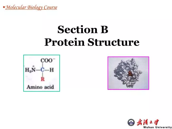
Molecular Biology Course. Section B Protein Structure. Cross with Biochemistry. Molecular Biology Course. B1 Amino acids: structure, side chains (charged, polar uncharged, nonpolar aliphatic, aromatic) B2 Protein structure and function:

E N D
Molecular Biology Course Section B Protein Structure
Cross with Biochemistry • Molecular Biology Course B1 Amino acids:structure, side chains (charged, polar uncharged, nonpolar aliphatic, aromatic) B2 Protein structure and function: Structure: size and shapes, primary secondary tertiary quaternary (prosthetic groups) ; Biological functions Structure & function: Domains, motif, and family B3 Protein analysis Purification Determinesequence, mass, and structure (X-ray crystallography and NMR)
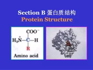
Protein structure COO- Ca H H3N R B1 Amino acids- basic structure Common structure of 19 AAs proline • a-carbon is chiral (asymmetric) except in glycine (R is H) 2. Amino acids are dipolar ions (zwitterions [兼性离子]) in aqueous solution and are amphoteric (兼性的同时有酸碱性或正负电荷的) 3. The side chains(R) differ in size, shape, charge and chemical reactivity 4. A few proteins contain nonstandard amino acids that are formed by post-translational modification of proline and lysine.
Protein structure B1 Amino acids- charged (5) Form salt bridges, are hydrophilic (亲水) 1. “Acidic” amino acids (2): containing additional carboxyl groups which are usually ionized aspartic acid(Asp, D,天冬氨酸) glutamic acid (Glu, E,谷氨酸)
Protein structure 2. “Basic” amino acids (3): containing positively charged groups e Lysine (Lys, K, 赖氨酸) Arginine(arg, R,精氨酸) d a guanidino group (胍基) Histidine(His, H,组氨酸) The imidazole group (咪唑基) has a pKa near neutrality. This group can be reversibly protonated under physiological conditions, which contribute to the catalytic mechanism of many enzymes.
Protein structure B1 Amino acids- polar uncharged (5) Contain groups that form hydrogen bonds with water, hydrophilic Serine(Ser, S,丝氨酸) Contain hydroxyl groups. Threonine(Thr, T,苏氨酸) Asparagine(Asn, N,天冬酰氨) Glutamine(Gln, Q,谷氨酰氨)
Protein structure Cysteine(Cys, C,半胱氨酸) has a thiol (巯醇) group, which is often oxidizes to cystine x-S-S-x
Protein structure B1 Amino acids- nonpolar aliphatic (7) (hydrophobic 疏水) Glycine (Gly, G,甘氨酸) Proline (Pro, P,脯氨酸): imino acid (亚氨基酸) Methionine(Met, M,甲硫氨酸): contains a sulfur atom
Protein structure Alkyl (烷基) side chains Alanine (Ala, A,丙氨酸) Leucine(Leu, L,亮氨酸) Isoleucine (Ile, I,异亮氨酸) Valine (Val, V,缬氨酸)
Protein structure B1 Amino acids- aromatic (3) Accounts for most of UV absorbance of proteins at 280 nm hydrophobic (疏水的) Phenylalanine (Phe, F,苯丙氨酸) Tyrosine (Tyr, Y,酪氨酸) Tryptophan (Trp, W,色氨酸)
B2 Protein structure and function • Molecular Biology Course Structure: size and shapes, primarysecondary tertiary quaternary, prosthetic groups (辅基,nonprotein molecules of conjugated proteins (结合蛋白) Domains, motif, and family Protein function
Protein structure B2 Protein structure -Sizes • A few thousands Daltons (x 103) to more than 5 million Daltons (x 106) • Some proteins contain bound nonprotein materials (prosthetic groups or other macromolecules), which accounts for the increased sizes and functionalities of the protein complexs.
Protein structure B2 Protein structure -Shapes Globular proteins: enzymes Complementary fit of a substrate molecule to the catalytic site on an enzyme molecule.
Protein structure Fibrous proteins: important structural proteins (silk fibroin, keratin in hair and wools ) Protofibril(初原纤维) microfibril(微管) Keratin(角蛋白) keratin in hair
Protein structure B2 Protein structure -Primary Polypeptidescontain N- and C- termini and are directional, usually ranging from 100-1500 aa Formation of a peptide bond (shaded in gray) in a dipeptide.
Protein structure N terminus C terminus Structure of the pentapeptide Ser-Gly-Tyr-Ala-Leu.
Protein structure B2 Protein structure -Secondary • a-helix • right-handed • 3.6 aa per turn • hydrogen bond • N-H···O=C A stereo, space-filling representation Collagen triple helix: three polypeptide intertwined
Protein structure b-sheet:hydrogen bonding of the pepetide bond N-H and C=O groups to the complementary groups of another section of the polypeptide chain Parallel b sheet: sections run in the same direction Antiparallel b sheet: sections run in the opposite direction A stereo, space-filling representation of the six-stranded antiparallel b sheet.
Protein structure B2 Protein structure -Tertiary The different sections of a-helix, b-sheet, other minor secondary structure and connecting loops of a polypeptide fold in three dimensions
Protein structure Noncovalent interaction between side chains that hold the tertiary structure together: van der Waals forces, hydrogen bonds, electrostatic salt bridges, hydrophobic interactions Covalent interaction: disulfide bonds Denaturation of protein by disruption of its 2o and 3o structure by heat and extremes of pH will lead to a random coil conformation
Protein structure B2 Protein structure -Quaternary Many proteins are composed of two or more polypeptide chains (subunits). These subunits may be identical or different. The same forces which stabilize tertiary structure hold these subunits together. This level of organization called quaternary structure. The quaternary structure of hemoglobin。 a1-yellow; b1-light blue; a2-green; b2-dark blue; heme-red back
Protein structure • Advantages of the quaternary structure: • It allows very large protein molecules to be made, such as tubulin。 • It can provide greater functionality to a protein by combining different activities into a single entity. • The interactions between the subunits can often be modified by binding of small molecules and lead to the allosteric effects seen in enzyme regulation
Protein structure B2 Protein structure - Prosthetic groups Covalently or noncovalently attached to many conjugated proteins, and give the proteins chemical functionality. Many are co-factors in enzyme reactions. Examples : heme groups in hemogobin (Figure)
Protein structure B2 Protein structure - Domains, motifs and families Domains:structurally independent units(tertiary) of a protein, being connected by sections with limited higher order structure within the same polypeptide. (Figure) They can also have specific function such as substrate binding
Protein structure • Structural motifs: • Groupings of secondary structural elements that frequently occur in globular proteins • Often have functional significance and represent the essential parts of binding or catalytic sites conserved among a protein family bab motif
Protein structure Protein families:structurally and functionally related proteins from different sources Motif The primary structures of c-type cytochromes from different organisms
Protein structure Domain The tertiary structures of the above c-type cytochromes Cys, Met and His side chains covalently linked to the heme to the protein Back
Protein structure B2 Protein functions • Enzymes: substrate binding, side chain in catalysis • Signaling: cell membrne • Transport and storage: hemoglobin transports oxygen • Structure and movement: collagen, keratin, tubulin in cytoskeleton, actin and myosin for muscle contraction • Nutrition: casein (酪蛋白) and ovalbumin(卵清蛋白) • Immunity: antibodies • Regulation: transcription factors
Protein structure B3 Protein analysis 1.Purification: to obtain enough pure sample for study 2. Sequencing: determine the primary structure of a pure protein sample 3. Mass determination: determine the molecular weight (MW) of an interested protein. 4. X-ray crystallography and NMR: determine the tertiary structure of a given sample.
Protein structure Protein purification • To purify the interested protein from other proteins and nonprotein molecules existing in the cells • An essential experimental step prior to study of any individual protein
Protein structure The principal properties of proteins used for purification • Size: gel filtration chromatography 2. Charge: ion-exchange chromatography, isoelectric focusing, electrophoresis 3.Hydrophobicity: hydrophobic interaction chromatography 4. Affinity: affinity chromatography 5. Recombinant techniques:involving DNA manipulation and making protein purification so easy
Protein structure • Gel filtration chromatography
Protein structure Bead:the matrix determine the size of the pore
Protein structure Ultracentrifugation:can be used to separate proteins according to their size and shape that determine their sedimentation rate. Very large protein complex.
Protein structure 2. Charge:ion-exchange chromatography, isoelectric focusing, electrophoresis Isoelectric point (pI): the pH at which the net surface charge of a protein is zero - + - + - + - + - + - - + + - + pH < pI pH=pI pH > pI
Protein structure Charge 1: Ion-exchange chromatography Sample mixture + + + Ion displacing Protein binding Column + anions Column + anions Column + proteins Purified protein
Protein structure Charge 2: Electrophoresis Protein migrate at different position depending on their net charge +
Protein structure Charge 3: Isoelectric focusing A protein will stop moving at position corresponding to its isoelectric point (pI) in a pH gradient gel.
Protein structure 3. Hydrophobicity: hydrophobic interaction chromatography Similar to ion-exchange chromatography except that column material contains aromatic or aliphatic alkyl groups
Protein structure 4. Affinity chromatography • Enzyme-substrate binding • Receptor-ligand binding • Antibody-antigen binding
Protein structure 5. Recombinant techniques: • Clone the interested protein encodinggene in an expression vector with a purification tag added at the 5’- or 3’ end of the gene • Protein overexpressionin a cell • Protein purification with affinity chromatography.
Protein structure Mass Determination Gel filtrationchromatography and SDS-PAGE • Comparing of the unknown protein with a proper standard • Popular SDS-PAGE: cheap and easy with a 5-10% error • SDS: sodium dodecyl sulfate, makes the proteins negatively charged and the overall charge of a protein is dependent on its mass.
Mass spectrometry: • Molecules are vaporized and ionized, and the degree of deflection (mass-dependent) of the ions in an electromagnetic field is measured • extremely accurate, but expensive • MALDI can measure the mass of proteins smaller than 100 KDa • Helpful to detect post-translational modification • Protein sequencing: relying on the protein data base
Protein structure Protein sequencing:Determine the primary structure of a protein • Specific enzyme/chemical cleavage: • Trypsin cleaves after lusine(K) or arginine (R) • V8 proteease cleaves after glutamic acid (E) • Cyanogen bromide cleaves after methionine (M) • Edaman degradation: • Performed in an automated protein sequencer • Determine thesequence of a polypeptide from N-terminal amino acid one by one. • Expensive and laborious
Protein structure Most protein sequences are deduced from the DNA/cDNA sequence Direct sequencing: determine the N-terminal sequences or some limited internal sequence construction of an oligonulceotide or antibody probe fishing the gene or cDNA
Protein structure X-ray crystallography and NMR Determing the tertiary structure (3-D) of a protein X-ray crystallography: • Measuring the pattern of diffraction of a beam of X-rays as it pass through a crystal. The first hand data obtained is electron density map, the crystal structure is then deduced. • A very powerful tool in understanding protein tertiary structure • Many proteins have been crystallized and analyzed
NMR • Measuring the relaxation of protons after they have been excited by radio frequencies in a strong magnetic field • Measure protein structure in liquid but not in crystal • Protein measured can not be larger than 30 KDa
Protein structure Protein Folding: chaperones are involved in vivo • The rapidly reversible formation of local secondary structure • Formation of domains through the cooperative aggregation of folding nulcei • Assembly of domains into a “molten” globule • Conformational adjustment of the monomer • Final conformational adjustment of the dimeric protein to form the native structure. back