Download

1 / 23
270 likes | 646 Views
Ch 4 Functional Anatomy of Prokaryotic and Eukaryotic Cells. Objectives. Compare and contrast the overall cell structure of prokaryotes and eukaryotes. Identify the three basic shapes of bacteria. Describe structure and function of the glycocalyx, flagella, axial filaments, fimbriae, and pili.

E N D
Objectives Compare and contrast the overall cell structure of prokaryotes and eukaryotes. Identify the three basic shapes of bacteria. Describe structure and function of the glycocalyx, flagella, axial filaments, fimbriae, and pili. Compare and contrast the cell walls of gram-positive bacteria, gram-negative bacteria, acid-fast bacteria, and mycoplasmas. Differentiate between protoplast, spheroplast, and L form. Describe the structure, chemistry, and functions of the prokaryotic plasma membrane. Identify the functions of the nuclear area, ribosomes, and inclusions. Describe the functions of endospores, sporulation, and endospore germination. What you should remember from Bio 31: Define organelle. Describe the functions of the nucleus, endoplasmic reticulum, ribosomes, Golgi complex, lysosomes, vacuoles, mitochondria, chloroplasts, peroxisomes. Explain endosymbiotic theory of eukaryotic evolution.
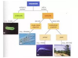
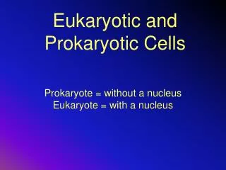
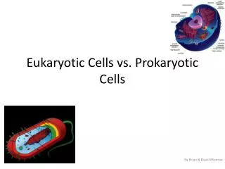
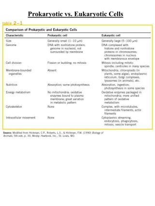
Common features: DNA and chromosomes Cell membrane Cytosol and Ribosomes Distinctive features: ? Comparing Prokaryotic and Eukaryotic Cells
Prokaryotes • One circular chromosome, not membrane bound • No histones • No membrane bound organelles • Peptidoglycan cell walls • Binary fission
Size, Shape, and Arrangement Average size: 0.2 -1.0 µm 2 - 8 µm Three basic shapes • Bacillus, -i • Coccus, -i • Spirals (Vibrio, Spirillum, Spirochete) Most monomorphic, some pleomorphic Variations in cell arrangements (esp. for cocci) Review Figs. 4.1, 4.2, and 4.4
Spiral Bacteria Figure 4.4
External Structures located outside of cell wall • Glycocalyx • Flagellum /-a • Axial filaments • Fimbria /-ae • Pilus /-i
Glycocalyx • Many bacteria secrete external surface layer composed of sticky polysaccharides, polypeptide, or both • Capsule: organized and firmly attached to cell wall • Slime layer: unorganized and loosely attached • Allows cells to attach key to biofilms • Prevents phagocytosis virulence factor • E.g.: B. anthracis, S. pneumoniae, S. mutans
Flagellum – Flagella • Anchored to wall and membrane • Number and placement determines ifatrichous, monotrichous, lophotrichous, amphitrichous, or peritrichous Fig 4.7
Flagellar Arrangement _______ ___________
Motility • Due to rotation of flagella • Mechanism of rotation: “Run and tumble” • Move toward or away from stimuli (taxis) • Stimuli (can be attractant or repellent) • Chemical (chemotaxis) • Light (phototaxis)
“Run and Tumble” Fig 4.9
Endoflagella In spirochetes Anchored at one end of a cell Rotation causes cell to move Fimbriae allow attachment Pili are used to transfer DNA from one cell to another Axial Filaments Fimbriae and Pili Fig 4.10
Cell Wall • Rigid for shape & protection prevents osmotic lysis • Consists of Peptidoglycan (murein) polymer of 2 monosaccharide subunits • N-acetylglucosamine (NAG) and • N-acetylmuramic acid (NAM) • Linked by polypeptides (forming peptide cross bridges) with tetrapeptide side chain attached to NAM • Fully permeable to ions, aa, and sugars (Gram positive cell wall may regulate movement of cations)
Thick layer of peptidoglycan Negatively charged teichoic acid on surface Thin peptidoglycan Outer membrane Periplasmic space Gram + Cell Wall Gram – Cell Wall
Gram-Positive Cell Walls • Teichoic acids • Lipoteichoic acid links to plasma membrane • Wall teichoic acid links to peptidoglycan • May regulate movement of cations • Polysaccharides provide antigenic variation
Gram-negative Cell Wall Lipid A of LPS acts as endotoxin; O polysaccharides are antigens for typing, e.g., E. coli O157:H7 Gram neg. bacteria are less sensitive to medications because outer membrane acts as additional barrier. LPS layer = outer layer of outer membrane (protein rich gel-like fluid) Fig 4.13
Gram Stain Mechanism • Crystal violet-iodine crystals form in cell. • Gram-positive • Alcohol dehydrates peptidoglycan • CV-I crystals do not leave • Gram-negative • Alcohol dissolves outer membrane and leaves holes in peptidoglycan. • CV-I washes out For further details and practical application see lab
Bacteria with No Cell Wall: Mycoplasmas • Instead, have cell membrane which incorporates cholesterol compounds (sterols), similar to eukaryotic cells • Cannot be detected by typical light microscopy This EM shows some typically pleomorphic mycoplasmas, in this case M. hyorhinis
Acid-fast Cell Walls • Genus Mycobacterium and Nocardia • mycolic acid (waxy lipid) covers thin peptidoglycan layer • Do not stain well with Gram stain use acid-fast stain
Damage to Cell Wall • Lysozyme (found in tears, mucus and saliva) digests disaccharide in peptidoglycan. • Penicillin inhibits peptide bridges in peptidoglycan.