Download
1 / 43
460 likes | 481 Views
LEUKEMIAS. By. Prof. Dr. Sameh Shamaa. Conditions characterised by abnormal proliferation of leucopoietic tissue and appearance of immature forms of white cells in the peripheral blood. Acute Leukemias

E N D
LEUKEMIAS By Prof. Dr. Sameh Shamaa
Conditions characterised by abnormal proliferation of leucopoietic tissue and appearance of immature forms of white cells in the peripheral blood.
Acute Leukemias Results from proliferation of young forms of leucocytes at a stage when they do not enter the circulation as readily as they do when more mature.
Classification: * acute lymphoblastic L. -----> more common * acute myeloblastic L. * acute monocytic L.
ACUTE LEUKEMIAS Def: Heterogenous group of neoplastic discases characterized. by proliferation of atypical elements which originates from the stem cells of the hematopoietic system. the uncontrolled and progressive proliferation of these cells lead to:- - Replacement of normal marrow - Invasion of peripheral blood -Infiltration of various organs and tissues
Etiology:- • Unknown, some factors seem to be implicated: - • A- Environmental factors:- • Ionizing radiation; mainly AML but ALL less important • Chemical substances; prolonged exposure to certain • substances (benzene, phenylbutazone, chloramphenicol, • anticancer drugs, alkylating agents, natulan) ---> incidence of leukemia mainly AML • Onset often preceeded by a state of bone marrow hypoplasia, peripheral pancytopenia • (preleukemic syndrome).
B-Genetic:- May act by facilitating envirnomental factors. C- Viruses:- Based on experiments with laboratory animal, has never proved humans. Typical products of viruses have been identified in some adult patients. With T.ALL:- HTLV (human T cell lymphotrophic virus) in T cell ALL and hairy cell leuk.
D- Immunological Factors: - Patient with acquired or congenital immune deficiency syndrome or subjected to prolonged immunosuppresive treatment has ↑ incidence of leukemia.
Differential Diagnosis : ------------------------------------------- D.D. between various forms of acute leukemia is mainly on cytomorphologic, cytochemical criteria rather than on clinical data, which are often superimposable. age: may accur at any age but in general: -A.L.L --- > the peak incidence in 1st 6 years of life. Uncommon afterage of 20 -A.M.L.--- >more commonly at slightly older age groups centering around the teen-age or adolescent period -A. Monocytic leuk. --->no specific age group.
Clinical Picture 1- Fever: moderate or high grade, usually irregular and shoots up when 2ry infection occur 2-rapidly developing marked asthenia. 3- rapidly developing severe anaemia ---> marked pallor. 4-severe bone ache allover the body 5-Severe sore throat 6- bleeding tendency from --->----- > gums. -------> skin --------> orifices --------->int. organs. 7-tender bones 8- joint pain (bleeding & infiltration) 9-L.N. enlargement in --->------> A.L.L. ---->A.M.L. ---->A. Monocytic L. 10-spleen & Liver enlargement. 11-Chloromas :- more in A.M.L. (subperiosteal tumor like masses) 12-C.N.S --->Focal Hge or infection of nerves or meningies.
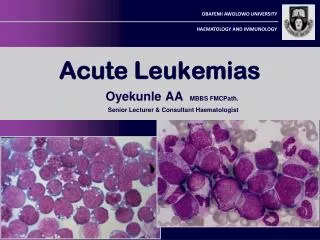
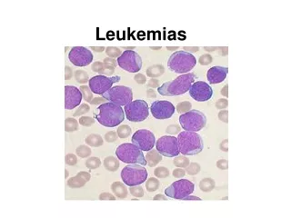
Blood Picture :- A.L.L : * R.B.Cs ---> severe form of normocytic ncrmochromic anemia occurs early. * W.B.cs ---> ( ) 10,000 --- 100,000 Or more but 40% are leukopenic - Lymphocytes are likely to be the predominant cells. - The diagnosis is established by detecting lymphoblastes in the peripheral smear. (N.B. Lymphoblastes : large cells with clear cytoplasm, prominant nucleus, definite nucleole. Diff. From mycloblastes :- absent specific granules of cytoplasm, -ve peroxidase stain -PAS +ve - Sudan Black -ve or weekly +ve * platelets : usually < 100.000 may be completetly absent.
2- Myeloblastic Leuk - W.B.Cs. count as A.L.L. - Leukopenia is as likely to occur as A.L.L - The cells are peroxidase +ve, PAS –ve, Sudan black +ve , acid phosphatase +ve - In 10 - 20% :Auer bodies are present rod like structures. in cytoplasm of------------> myeloblast -------------> monoblast 3- Monocytic Leukemia: - monoblast in the peripheral blood. Stain PAS -ve, Sudan B +ve, Peroxidase -ve acid phosphatase +ve
Bone marrow aspiration: Massive proliferation of blast cells even when leukopenia exist. * A.L.L-----> lymphoblast * A.M.L---->myeloblast *A. monocytic----> monoblasts and monocytes. *Radiology :- -Sketetat involvement in almost all children and 50% of dults. * diffuse osteoprosis * periosteal elevation * osteolytic lesions * radiolucent metaphyseal bands.
Diff- diagnosis :- The combination of * anemia * thrombocytopenia * bone marrow prolif. è primitive white cells is found only in leukemia.
From other causes ofsorethroat + Fever as:- • * vincent angina. • *diphtheria. • *infectious, mononucleosis. • *aplastic anemia. • *agranulocytosis. • (2) from other causes of parpura :- • * I.T.P • *Aplastic anemia.
(3) Lymphadenopathy + splenomegally:- * infective mononucleosis *H.D. .N.H.L. (by blood picture).. (4) Lymphocytosis :in :. * whooping caugh *infective lyrmphocytosis. (white cells mature, R.B.Cs and platelets are normal). (5) Rheumtic Fever
The FAB classificationSubtyping of the various forms of ALL used to be done according to the French-American-British (FAB) classification,[18] which was used for all acute leukemias (including acute myelogenous leukemia, AML). ALL-L1: small uniform cells ALL-L2: large varied cells ALL-L3: large varied cells with vacuoles (bubble-like features) Each subtype is then further classified by determining the surface markers of the abnormal lymphocytes, called immunophenotyping. There are 2 main immunologic types: pre-B cell and pre-T cell. The mature B-cell ALL (L3) is now classified as Burkitt's lymphoma/leukemia. Subtyping helps determine the prognosis and most appropriate treatment in treating ALL
WHO proposed classification of acute lymphoblastic leukemia • The recent WHO International panel on ALL recommends that the FAB classification be abandoned, since the morphological classification has no clinical or prognostic relevance. It instead advocates the use of the immunophenotypic classification mentioned below. • 1- Acute lymphoblastic leukemia/lymphoma Synonyms:Former Fab L1/L2 • i. Precursor B acute lymphoblastic leukemia/lymphoma. Cytogenetic subtypes:[19] • t(12;21)(p12,q22) TEL/AML-1 • t(1;19)(q23;p13) PBX/E2A • t(9;22)(q34;q11) ABL/BCR • T(V,11)(V;q23) V/MLL • ii. Precursor T acute lymphoblastic leukemia/lymphoma • 2- Burkitt's leukemia/lymphoma Synonyms:Former FAB L3 • 3- Biphenotypic acute leukemia
Treatment:- Combination chemotherapy in order to :- * obtain synergestic action * minimize side effects. * attacks leukemic cells in different phases of mitosis. * delay the onset of resistance of the malignant cells.
Acute L. Leuk.effective drugs are 1- vincristine-----> arrest cell mitosis 2- predinsone ----> Lyrmpholysis 3-6.M.P. ----> inhibit DNA synthesis. 4-Methotrexate ----> inhibit RNA and protein synthesis 5-Doxorubich (adriamycin)----> inhibit DNA synthesis 6-L- asparaginase
Intensification Intensification uses high doses of intravenous multidrug chemotherapy to further reduce tumor burden. Since ALL cells sometimes penetrate the Central Nervous System (CNS), most protocols include delivery of chemotherapy into the CNS fluid (termed intrathecal chemotherapy). Some centers deliver the drug through Ommaya reservoir (a device surgically placed under the scalp and used to deliver drugs to the CNS fluid and to extract CNS fluid for various tests). Other centers would perform multiple lumbar punctures as needed for testing and treatment delivery. Typical intensification protocols use vincristine, cyclophosphamide, cytarabine, daunorubicin, etoposide, thioguanine or mercaptopurine given as blocks in different combinations. For CNS protection, intrathecal methotrexate or cytarabine is usually used combined with or without cranio-spinal irradiation (the use of radiation therapy to the head and spine). Central nervous system relapse is treated with intrathecal administration of hydrocortisone, methotrexate, and cytarabine.
Maintenance therapy The aim of maintenance therapy is to kill any residual cell that was not killed by remission induction, and intensification regimens. Although such cells are few, they will cause relapse if not eradicated. For this purpose, daily oral mercaptopurine, once weekly oral methotrexate, once monthly 5-day course of intravenous vincristine and oral corticosteroids are usually used. The length of maintenance therapy is 3 years for boys, 2 years for girls and adults.
These drugs must be used sequentially and in combinations:- a-Induction therapy:- * To obtain apparent clinical & hematological remission * 2 or 3 drugs used - Vincristine 1.4 mg/m2 I.V. once weekly for 4 weeks... - prednisone lmg/kg body weight P.O. daily - L. asparaginaseor Adriamycin
b- Consolidation theraby:- *to decrease total no. of residual malignant cells to 10G cells or less. * e.g. Methotrexate : 15 mg/m2I.M. daily For 3 - 5 days.. followed by cystarabine 100 mg/ m2 I.V. twice daily for 3-5 days.
c- C.N.S. prophylaxis by * cranial irradiation1800 2400 R * Intra thecal (l.T.) Methotrexate in mg/ m2 for 5 injection. (twice weekly).
d- Prolonged maintenance :~ * 2-3 years * Methotrexate 15 mg/ m2once or twice weekly I.M. * 6M.P 1-2.5 mg/kg daily P.O. *Cyclophosphamide: 200mg/ m2 p.o. weekly.
Acute Non Lymphoblastic L. -The initial objectives is the induction of a CR. (reduction of marrow blasts to less than 5%, increase in neutrophils in the peripheral blood to l.5x 106 /L. or more and of platelets to 100x 106/L. or more.). -Then consolidation therapy follows, for which identical drugs are used (the aim is further reduction of leukemic cells). - Both in induction and consolidation, high dosage of drugs are given to produce temporary marrow aplasia.
After the marrow have recovered from consolidation therapy, maintenance therapy is given at monthly intervals usually for 2-3 years, aiming for a further stepwise reduction of remaining leukemic cells without making the marrow splastic • -The cornerstone of remission induction and consolidation chemotherapy is a combination of cytosine-arabinoside and daunorubicin resulting in 50% CR rates. • -Drugs used for maintenance: cytosine-arabinoside, 6-thioguanine, cyclophosphamide and daunarubicin. • However the value of maintenance treatment is marjinal.
complications :- (1)Severe bone ache or massive C.N.S infiltration. ---- > Local irradiation (2)C.N.S. involvement: ----> intrathecal Mtx. 12 mg I.T./3 days + cranial irradiation (3)Fever. (4)Hge. (6)Hyperuricemia
Prognosis (1) A.L.L ----->C.R. ----> 90% of children. ----> 60% of adults. ----> Cure in 70% of children. and 35% of adults. Allogenic bone marrow transplantation is the only formoftherapy for patients with drug -resistant disease (2) A.N.L.L.----->C.R is up to 50% - only about 25% remain disease free after 5 years and can expect to be cured - However, bone marrow transplantation if preformed during First remission can improve survival and it has been suggested to be preformed in all patients younger than 50 years and who have available HLA identical donor
[edit] Prognosis The survival rate has improved from zero four decades ago, to 20-75 percent currently, largely due to clinical trials and improvements in bone marrow transplantation (BMT) and stem cell transplantation (SCT) technology. Five-year survival rates evaluate older, not current, treatments. New drugs, and matching treatment to the genetic characteristics of the blast cells, may improve those rates. The prognosis for ALL differs between individuals depending on a variety of factors: Sex: females tend to fare better than males. Ethnicity: Caucasians are more likely to develop acute leukemia than African-Americans, Asians and Hispanics and tend to have a better prognosis than non-Caucasians. Age at diagnosis: children between 1–10 years of age are most likely to develop ALL and to be cured of it. Cases in older patients are more likely to result from chromosomal abnormalities (e.g. the Philadelphia chromosome) that make treatment more difficult and prognoses poorer. White blood cell count at diagnosis of less than 50,000/µl Cancer spread into the Central_nervous_system (brain or spinal cord) has worse outcomes. Morphological, immunological, and genetic subtypes Patient's response to initial treatment Genetic disorders such as Down's Syndrome
Cytogenetics, the study of characteristic large changes in the chromosomes of cancer cells, is an important predictor of outcome.[13] Some cytogenetic subtypes have a worse prognosis than others