1 / 1

FIGURE 1 . Confocal micrographs of samples from 5 culture-negative,

FIGURE 1 . Confocal micrographs of samples from 5 culture-negative, Ibis-positive cases in which the tissues were evaluated with FISH probes (pink) integrated into the

kali
Download Presentation

FIGURE 1 . Confocal micrographs of samples from 5 culture-negative,

An Image/Link below is provided (as is) to download presentation Download Policy: Content on the Website is provided to you AS IS for your information and personal use and may not be sold / licensed / shared on other websites without getting consent from its author. Content is provided to you AS IS for your information and personal use only. Download presentation by click this link. While downloading, if for some reason you are not able to download a presentation, the publisher may have deleted the file from their server. During download, if you can't get a presentation, the file might be deleted by the publisher.

E N D

Presentation Transcript


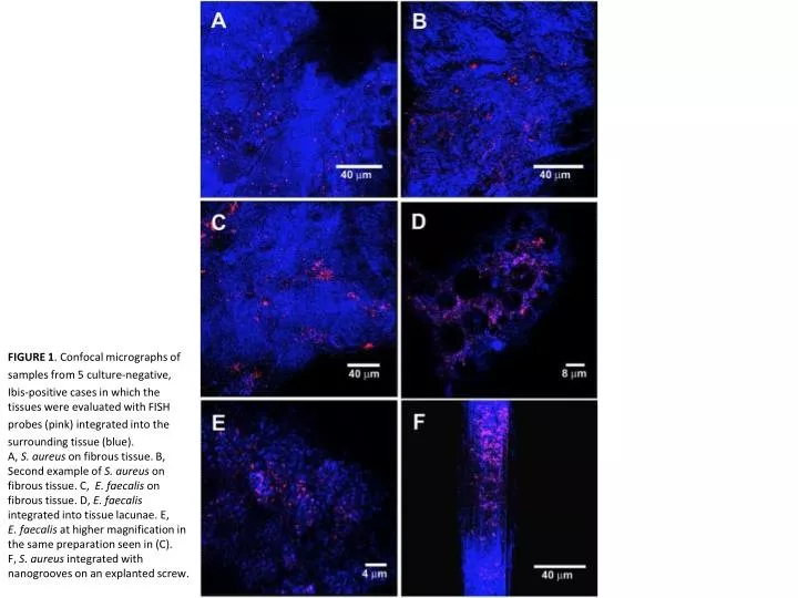
  1. FIGURE 1. Confocal micrographs of samples from 5 culture-negative, Ibis-positive cases in which the tissues were evaluated with FISH probes (pink) integrated into the surrounding tissue (blue). A, S. aureuson fibrous tissue. B, Second example of S. aureus on fibrous tissue. C, E. faecalison fibrous tissue. D, E. faecalisintegrated into tissue lacunae. E, E. faecalis at higher magnification in the same preparation seen in (C). F, S. aureusintegrated with nanogrooves on an explanted screw.

More Related