Download
1 / 84
860 likes | 1.07k Views
THORACIC WALL CARDIOVASCULAR SYSTEM. The Two Fridas 1939 by Frida Kahlo. 01. 10. 2013. Kaan Yücel M.D., Ph.D. 1. THORAX. the part between the neck and the abdomen. Chest X-ray. 1.1. REGIONS/T E RMS. Thoracic cavity cavity between neck and abdomen

E N D
THORACIC WALL CARDIOVASCULAR SYSTEM TheTwoFridas1939 byFridaKahlo 01. 10. 2013 Kaan Yücel M.D., Ph.D.
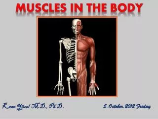
1. THORAX the part between the neck and the abdomen Chest X-ray
1.1. REGIONS/T ERMS Thoracic cavity cavity between neck and abdomen protected by the thoracic wall. Thoracic wall bounds the thoracic cavity. formed by the skin, bones, fasciae, and muscles. Thoracic cage bony portion of the thoracic wall thoracic skeleton
1.2. SURFACES OF THE THORAX STERNUM & COSTAL CARTILAGES anteriorly 12 THORACIC VERTEBRAE & POST. RIBS posteriorly RIBS & INTERCOSTAL SPACES laterally Posterior surface 12 thoracic vertebræ& posterior parts of the ribs Anterior surface sternum &costal cartilages Lateral surfaces ribs, separated by the intercostal spaces
1.3. BOUNDARIES OF THE THORAX • Superior • Jugular notch • Sternoclavicular joint • Superiorborder of clavicle • Acromion • Spinousprocesses of C7 • Inferior • Xiphoid process • Costal arch • 12th and 11th ribs • VertebraT12
1.4. CONTENTS OF THE THORAX Organs of the cardiovascular, respiratory, digestive, reproductive, immune, and nervous systems
2. THORACIC WALL • thoracic cage (skeleton) • muscles between the ribs • skin • subcutaneous tissue • muscles, and fascia covering its anterolateral aspect. • The mammary glands of the breasts lie within the subcutaneous tissue of the thoracic wall.
2.1. Functions of the thoracic wall • Protectsvital thoracic and abdominal organs • Resiststhe negative (sub-atmospheric) internal pressures generated by the elastic recoil of the lungs and inspiratory movements. • Providesattachmentfor and support the weight of the upper limbs. • Provides theoriginsof many of the muscles that move and maintain the position of the upper limbs relative to the trunk. • Provides attachments for muscles of the abdomen, neck, back, and respiration.
3. Skeleton of THEThoracic Wall • 12 pairs of ribs and associated costal cartilages • 12 thoracic vertebrae and the intervertebral (IV) discs interposed between them • Sternum
4. THORACIC APERTURES ‘Thoracic inlet’ ‘Thoracicoutlet’
4.1. Superior thoracic aperture “doorway” between the thoracic cavity and the neck and upper limb • bounded: • Posteriorly vertebra T1 • Laterally 1st pair of ribs and their costal cartilages • Anteriorlysuperior border of the manubrium • Trachea • Esophagus • nerves, and vessels that supply and drain the head, neck, and upper limbs.
4.2. Inferior thoracic aperture By closing the inferior thoracic aperture, the diaphragm separates the thoracic and abdominal cavities almost completely. • bounded: • Posteriorly12th thoracic vertebra • Posterolaterally11th and 12th pairs of ribs • Anterolaterallyjoined costal cartilages of ribs 7-10costal margins • Anteriorlyxiphisternal joint
5. MOVEMENTS OF THE THORACIC WALL One of the principal functions of the thoracic wall and the diaphragm is to alter the volume of the thorax and thereby move air in and out of the lungs. During breathing, the dimensions of the thorax change in vertical, lateral, and A-P directions. Diaphragm contracts Depression Diaphragmrelaxes Elevation (duringpassiveexpiration) Elevation&depression of the ribs
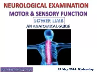
Dermatomes Skin areasupplied by a segment of the spinal cord Through its posterior ramusand the lateral and anterior cutaneous branches of its anterior ramus, most thoracic spinal nerves (T2-T12) supply a strip-like dermatome of the trunkextending from the posterior median line to the anterior median line. T2- Sternal angle T4- Nipple T6- Xiphoid process T8- Costal arch T10-Umbliculus T12-Midpoint between umbilicus and symphysis pubis
2. breasts Reproduction, backpain Aesthetics, and breast cancer Mammaryglands &associated skin -connective tissues. modified sweat glands in the superficial fascia anterior to the pectoral muscles and the anterior thoracic wall.
2. breasts Mammaryglands: Seriesof ducts and associated secretory lobules. Form15 to 20 lactiferous ductsopen nipple. Nippleis surrounded by a circular pigmented area of skin areola(L. small area).
Female Breasts NON-LACTING WOMEN – PREDOMINANT COMPONENT: FAT LACTING WOMEN- PREDOMINANT COMPONENT: GLANDULAR TISSUE The breast rests on a bed extends transversely from lateral border of the sternum mid-axillary line vertically from the 2nd through 6th ribs
Lymphaticdrainage of thebreast 75% (lateralbreastquadrants) Axillarylymphnodes Most of theremaining (medialbreastquadrants) parasternallymphnodesortotheoppositebreast Lymphfrominferiorquadrantsmaypassdeeplytoabdominallymphnodes.
Axillarylymphnodes Parasternallymphnodes Bronchomediastinallymphtrunks Clavicularlymphnodes Subclavianlypmhatictrunk Right orleftvenousangle .
3. HEART (1)7.3 million • Trapezoidalin A-P dimensions • Tipped-over pyramid in 3-D • crucial organ of the human body 30%
Leftheart (Pumping) well- oxygenated(arterial) blood fromthelungs pulmonaryveins leftatriumleftventricle aorta the body Right heart (Suction) poorly- oxygenated(venous) blood fromthe body superior vena cava & inferior vena cava rightatriumrightventricle pulmonaryarteries lungs
The four chambers of the heart right and left atria & right and left ventricles Ventricles Dischargingchambers Atrium – pluralatria Receivingchambers cardiac cycle • Ventricularfilling (diastole) • Ventricularemptying (systole) Blood pressure 120-80 mm/Hg
The fibrous skeleton of the heart • Keeps the orifices of the AV &semilunar valves patent • prevents them from being overly distended by an increased volume of blood. • Provides attachments for the valves & myocardium. • Forms an electrical «insulator» • separating impulses of the atria & ventricles they contract independently • surrounding and providing passage for the initial part of the AV bundle
Sulci/Grooves in the heart coronary sulcus (atrioventricular groove) betweenatrium & ventricles anterior &posterior interventricular (IV) sulci (grooves) betweenright and left ventricles
Apex &base of the heart apex located inferiorly & base located superiorly • Apexprojectsforward, downwardandtotheleft • Base faces in a posteriordirection
The four surfaces of the heart • Anterior (sternocostal) surface • mostly of right ventricle • someof the right atrium on the right • some of the left ventricle on the left • Diaphragmatic (inferior) surface • formed mainly by the left ventricle • partly by the right ventricle • related to central tendon of diaphragm. • Right pulmonary surface • formed by the right atrium. • Left pulmonary surface • left ventricle &a portion of left atrium.
RIGHT ATRIUM forms the right border of the heart • Receives venous blood from the SVC, IVC, and coronary sinus. • Through the right atrioventricular orifice, • discharges the poorly oxygenated blood it has received • into the right ventricle.
RIGHT VENTRICLE • forms • largest part of the anterior surface of the heart • a small part of the diaphragmatic surface • almost the entire inferior border of the heart.
interventricular septum (IVS) obliquely placed partition between the right and left ventricles, forming part of the walls of each muscular and membranous parts • Bulgesinto the cavity of the right ventricle. • Superiorly and posteriorly, a thin membrane, forms the much smaller membranous part of the IVS.
LEFT ATRIUM forms most of the base of the heart • right and left pulmonary veins enter here. • Tubular, muscular left auricle, • Itswall trabeculated with pectinatemuscles. • A semilunar depression in the interatrial septum • Floor of the oval fossa • surrounding ridge • Valveof the oval fossa
LEFT VENTRICLE forms the apex of the heart, left (pulmonary) surface & border, most of the diaphragmatic surface. • Comparedtotherightventricle • Walls 2-3 times thicker • Trabeculaecarneaefiner and more numerous • Cavity longer • Anterior &posterior papillary muscles larger
aortic valve • semilunarvalve • between the left ventricle &ascending aorta • obliquely placed.
mitral valve double-leaflet mitral valve • Guardsthe left AV orifice. • Has two cusps, anterior and posterior.
6. SEMILUNAR VALVES Semilunarcusps of thepulmonaryvalveanterior-right-left Seminularcusps of theaorticvalveposterior-right-left concavewhen viewed superiorly notendinouscords to support
VASCULATURE OF THE HEART coronary arteries & cardiac veins • embedded in fat • course across the surface of the heart just deep to the epicardium.
Arterial Supply of the Heart coronary arteries first branches of the aorta supply the myocardium and epicardium Anastomoses between the branches of the coronary arteries exist, which enables the development of the collateral circulation.
STIMULATING, CONDUCTING, & REGULATINGSYSTEMS OF HEART 1.sinuatrial(SA) node initiates the heartbeat &coordinates contractions of the four heart chambers • 2.atrioventricular (AV) node • 3.bundles • highly specialized conducting fibers for conducting impulsesrapidly to different areas of the heart • Propagation of theimpluse • Simultaneouscontraction of thecardiacstriatedmusclecells
sinuatrial (SA) node pacemaker of the heart • @junction of the SVC&right atrium • near tothesuperior end of thesulcus terminalis
sinuatrial (SA) node pacemaker of the heart stimulated by sympathetic division of the autonomic nervous system to accelerate the heart rate inhibited by parasympathetic division to return to or approach its basal rate.
atrioventricular(AV) node a smaller collection of nodal tissue than the SA node • in the posteroinferior region of the interatrialseptum • near the opening of the coronary sinus
JOURNEY OF THE SIGNAL • Generated @ SA node • Passedthrough the walls of the right atrium • Propagetedbythecardiacmuscle • Signalpassedfrom SA nodeto AV node • Distributed totheventriclesthroughthe AV bundle
BUNDLES AV bundle the only bridge between the atrial and ventricular myocardium • passes from the AV node • through the fibrous skeleton of the heart and along the membranous part of the IVS. • @ junction of membranous & muscularparts of the IVS • dividesinto : rightbundle & leftbundle.
BUNDLES right and left bundles proceed on each side of the muscular IVS deep to the endocardium then ramify into subendocardialbranches (Purkinje fibers) extend into the walls of the respective ventricles.
Innervationof the Heart autonomicnervoussystem, cardiacplexus Cardiacplexus posterior to the ascending aorta and bifurcation of the pulmonary trunk
Innervationof the Heart autonomicnervoussystem, cardiacplexus • Parasympatheticsupply • presynaptic fibers of the vagusnerves • Slows the heart rate • Reducesthe force of the contraction • Constrictsthe coronary arteriessaving energy
Innervationof the Heart • sympathetic supply • presynaptic fibers • cell bodies in the intermediolateral cell columns (IMLs) of the superior 5or 6thoracic segments • postsynaptic sympathetic fibers • cell bodies in the cervical and superior thoracic paravertebral ganglia of the sympathetic trunks. • causes increased heart rate • increased impulse conduction, increasedforce of contraction, • increased blood flow through the coronary vessels increased activity.
9. SEPTAL DEFECTS Atrial Septal Defects (ASD) congenital anomaly of the interatrialseptum a hole betweenthetwoatria Results in Whathappens? enlargement of right atrium & ventricle dilation of the pulmonary trunk Oxygenatedbloodfromthelungs Leftatrium Right atrium Moreblood in therightheart
VentricularSeptalDefects (VSD) rank first on all lists of cardiac defects membranous part of the IVS common site of VSDs Results in Whathappens? in pulmonary blood flow severe pulmonary disease (hypertension) cardiac failure Oxygenatedbloodfromtheventricles Leftventricle Right ventricle
10. VALVULAR HEART DISEASES • Disturb pumping efficiency of the heart. • Stenosis(narrowing) or insufficiency • Both result in an increased workload for the heart. • Valvuloplastyrepairingtheheartvalves
Mitral Valve Insufficiency • Scarring and shortening of the cusps results in insufficiency • Restricts the outflow of the left ventricle • Leadsto the hypertrophy of the myocardium • During ventricular systole, blood regurgitates back to the left atrium • A hurt murmur will be heard.