Download
1 / 88
930 likes | 1.59k Views
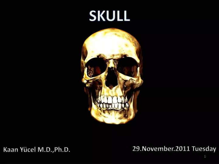
SKULL. 29.November.2011 Tuesday. Kaan Yücel M.D.,Ph.D. The cranium (skull) is the skeleton of the head. A series of bones form its two parts: Neurocranium Viscerocranium (Facial bones ).

E N D
SKULL 29.November.2011 Tuesday Kaan Yücel M.D.,Ph.D.
The cranium (skull) is the skeleton of the head. A series of bones form its two parts: Neurocranium Viscerocranium (Facial bones)
The neurocraniumis the bony case of the brain and its membranous coverings, the cranial meninges. It also contains proximal parts of the cranial nerves and the vasculature of the brain. The neurocranium formed by 8 bones: 4 singular bones (Frontal, ethmoidal, sphenoidal, and occipital) 2 sets of bones bilateral pairs (Temporal and parietal)
The neurocranium has a dome-like roof, calvaria(skullcap), and a floor or cranial base (basicranium). The bones making the calvaria are primarily flat bones (frontal, parietal, and occipital). The bones contributing to the cranial base are primarily irregular bones with substantial flat portions (sphenoidal and temporal).
The bones contributing to the cranial base are primarily irregular bones with substantial flat portions (sphenoidal and temporal).
The viscerocranium (facial skeleton) comprises the facial bones. • The viscerocranium forms the anterior part of the cranium and consists of the bones surrounding • mouth(upper and lower jaws) • nose/nasal cavity • most of the orbits (eye sockets or orbital cavities).
The viscerocranium consists of 15 irregular bones: 3 singular bones (mandible, ethmoid, and vomer) 6 bones bilateral pairs (maxillae; inferior nasal conchae; and zygomatic, palatine, nasal, and lacrimal bones).
The maxillae and mandible house the teeth—that is, they provide the sockets and supporting bone for the maxillary and mandibular teeth. • The maxillae contribute the greatest part of the upper facial skeleton, forming the skeleton of the upper jaw. • The mandible forms the skeleton of the lower jaw.
Several bones of the cranium (frontal, temporal, sphenoid, and ethmoid bones) are pneumatized bones, which contain air spaces (air cells or large sinuses), presumably to decrease their weight. The total volume of the air spaces in these bones increases with age.
Ossa Cranii Frontal Bone Resembles a cockle-shell in form. Consists of two portions: Squama, corresponding with the region of the forehead Orbital portion, enters into the formation of the roofs of the orbital and nasal cavities.
Frontal Bone • Articulates inferiorly with the nasal and zygomatic bones. • Also articulates with the lacrimal, ethmoid, and sphenoids.
Frontal Bone A horizontal portion of bone (orbital part) forms both the roof of the orbit and part of the floor of the anterior part of the cranial cavity. The intersection of the frontal and the nasal bones is thenasion (L. nasus, nose); distinctly depressed area (bridge of nose).
Frontal Bone The supra-orbital margin of the frontal bone, the angular boundary between the squamous and the orbital parts, has a supra-orbital foramen or notch. The internal surface of the squamafrontalis of the frontal bone is concave Presents a ridge, frontal crest; median bony extension of the frontal bone.
Frontal Bone Nasal process is the downward projection of the nasal part of the frontal bone which terminates as the nasal spine.
Parietal Bones • Form the roof of the cranium. • Irregularly quadrilateral in form. • The external surface is convex,, and marked near the center by parietal eminence (tuber parietale). • Crossing the middle of the bone in an arched direction are two curved lines, superior and inferior temporal lines.
Temporal Bones Situated at the sides and base of the skull. Each consists of 5 parts, squama, petrous, mastoid, and tympanic parts, and t styloid process.
Temporal Bones The temporal fossais bounded: Superiorly and posteriorly by the superior and inferior temporal lines, Anteriorly by the frontal and zygomatic bones Inferiorly by the zygomatic arch.
Temporal Bones The temporal bone contains, in its interior, the essential parts of the organ of hearing and the organ of equilibrium.
Temporal Bones Its inferior surface contains an oval depression which is called the mandibular fossa, articulates with the mandible.
Sphenoid Bone Situated at the base of the skull in front of the temporals and basilar part of the occipital. Somewhat resembles a bat with its wings extended, and is divided into a median portion or body, two greatand two small wingsextending outward from the sides of the body, and two pterygoid processes which project from it below.
Sphenoid Bone • The sella turcica (L. Turkish saddle) is the saddle-like bony formation on the upper surface of the body of the sphenoid. • Composed of 3 parts: • • Tuberculumsellae(Hornof saddle) • •Hypophysialfossa (Pituitaryfossa) • •Dorsumsellae(Backof saddle)
Sphenoid Bone • On each side of the body of the sphenoid, a crescent of four foramina perforate the roots of the cerebral surfaces of the greater wings of the sphenoids: • Superior orbital fissure • Foramen rotundum (round foramen) • Foramen ovale (oval foramen) • Foramen spinosum (spinous foramen)
Occipital Bone Situated at the back and lower part of the cranium. Pierced by a large oval aperture, foramen magnum, through which the cranial cavity communicates with the vertebral canal.
Occipital Bone The cranial base is formed posteriorly by the occipital bone, which articulates with the sphenoid bone anteriorly.
Occipital Bone The hypoglossal canal for the hypoglossal nerve (CN XII) is superior to the anterolateral margin of the foramen magnum.
Ethmoid Bone Situated at the anterior part of the base of the cranium, between the two orbits, at the roof of the nose, and contributes to each of these cavities.
Ethmoid Bone • Consists of 4 parts: • A horizontal or cribriform plate, forming part of the base of the cranium; • A perpendicular plate, constituting part of the nasal septum; • Its numerous tiny foramina transmit the olfactory nerves (CN I).
Cranial Fossas • Anterior cranial fossa • Shallowest of the three cranial fossae. • Formed by the frontal bone anteriorly, the ethmoid bone in the middle, and the body and lesser wings of the sphenoid posteriorly.
Middle cranial fossa • Butterfly-shaped • Has a central part composed of the sella turcica on the body of the sphenoid.
Posterior cranial fossa • Largest and deepest of the three cranial fossae • Formed mostly by the occipital bone, but the dorsum sellae of the sphenoid marks its anterior boundary centrally and the petrous and mastoid parts of the temporal bones contribute its anterolateral “walls.”
L. maxilla "upper jaw," diminutive of mala "jaw, cheekbone." Largest bones of the face, excepting the mandible Form, by their union, the whole of the upper jaw.
Their alveolar processes include the tooth sockets (alveoli) and constitute the supporting bone for the maxillary teeth.
The two maxillae are united at the intermaxillary suture in the median plane. They have a broad connection with the zygomatic bones laterally.
Each maxilla assists in forming the boundaries of 3 cavities: Roof of the mouth Floor & lateral wall of the nose Floor of the orbit
It also enters into the formation of two fossæ, the infratemporaland pterygopalatine, and two fissures, the inferior orbitaland pterygomaxillary.
Superiorly,each maxilla contributes to the inferior and medial rims of the orbit. Laterally, the zygomatic process of each maxilla articulates with the zygomatic bone. Medially, the frontal process of each maxilla articulates with the frontal bone. Inferiorly, the part of each maxilla, lateral to the opening of the nasal cavity, is the body of maxilla.
The body of the maxillais large and roughly pyramidal in shape. Its interior is hollowed out by the maxillary paranasal air sinus. • Upper (orbital) surface of the body occupies the floor of the orbit. • Anterior surface forms the curved external surface of the upper jaw. • Posterior (infratemporal) surface provides the anterior wall of the infratemporal fossa. • Medial (nasal) surface is a major structure component in the wall of the nasal cavity.
Upper (orbital) surface of the body Medial (nasal) surface Anterior surface Posterior (infratemporal) surface
Maxillary tuberosity, located behind the last molar tooth. More medially the lower part of the infratemporal surface articulates with the pyramidal process of the palatine bone.
The four maxillary processes: Zygomatic process Frontal process Palatine process Alveolar process
Above the incisor teeth, the anterior (oral) surface has shallow depression in the midline, termed the incisive fossa. It receives the lower openings of the two incisive canals which communicate above with the corresponding halves of the nasal cavity.
Further laterally is the deeper canine fossa which is separated from the incisive fossa by the canine eminence produced by the root of the canine tooth.
Above the canine fossa is the infraorbital foramen. The anterior surface ends medially at the anterior nasal aperture. At the inferior margin of this aperture the maxillae of the two sides form a median projection, the anterior nasal spine.
The bony palate (the term hard palate is used to describe the bony palate plus its covering of mucous membrane; the bony palate thus forms the skeleton of the hard palate) provides the floor of the nasal cavity and the roof of the mouth. Its anterior three-quarters are formed by the palatine process of the maxillae and its posterior one-quarter by the horizontal plates of the palatine bones.
2 sutures cross the palate: Median suture between the elements of the right and left sides Transverse suture between the palatine processes of the maxillae and the horizontal plates of the palatines. The four bony parts of the hard palate — the paired maxillary palatine processes (1) and the paired horizontal plates of the palatine bones (2) — form a cross-shaped suture (3), also known as the intermaxillary suture. This suture marks the juncture of each side of the hard palate; and the transverse suture (4), the junction of the maxillary and palatine bones.