Download
1 / 57
570 likes | 674 Views
This presentation will probably involve audience discussion, which will create action items. Use PowerPoint to keep track of these action items during your presentation In Slide Show, click on the right mouse button Select “Meeting Minder” Select the “Action Items” tab

E N D
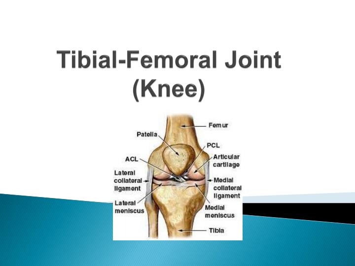
This presentation will probably involve audience discussion, which will create action items. Use PowerPoint to keep track of these action items during your presentation • In Slide Show, click on the right mouse button • Select “Meeting Minder” • Select the “Action Items” tab • Type in action items as they come up • Click OK to dismiss this box • This will automatically create an Action Item slide at the end of your presentation with your points entered. Tibial-Femoral Joint (Knee)
Movements of the Knee • Hinge joint, that is stabilized by four ligaments and very strong musculature. • Extension • Flexion • Rotation (very slightly)
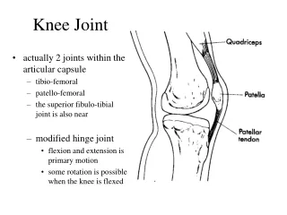
Bones • Fibula • Tibia • Patella • Largest sessmoid bone in the human body • Patella slides up and down as the knee flexes and articulates with the femur. (patellar-femoral joint) • Femur
Cartilage • Articular Cartilage • On the distal end of the femur • Posterior side of patella • Meniscus • Sits on the tibial plateau • Its job is to cushion for shock absorption and aid in the stabilization of the joint • Thicker on the sides and thinner in the middle (bowl shaped)
Ligaments • Four Primary Ligaments • Medial Collateral Ligament (MCL) • Tibial Collateral Ligament • Lateral Collateral Ligament (LCL) • Fibular Collateral Ligament • Anterior Cruciate Ligament (ACL) • Limits anterior displacement of the tibia • Posterior Cruciate Ligament (PCL) • Limits posterior displacement of the tibia
Muscles • Quadriceps • Four separate muscles • Rectus Femoris • Vastus Lateralis • Vastus Intermedius • Vastus Medialis • Extends the knee • Hamstrings • Three separate muscles • Biceps Femoris • Semimembranosus • Semitendinosus • Flexes the knee
Bursa Sacs • Fluid filled sacs • Aid in protection for direct trauma • May become inflammed or injured after a direct blow to the knee.
Prepatellar Bursa Pretibial Bursa Infrapatellar Bursa
Knee Ligament Injuries • Knee ligament injuries can happen individually or in combination. • How many ligaments depends on the types of forces: • Valgus force- a direct blow from the lateral side affecting medial side structures • Varus force- a direct blow from the medial side, affecting lateral side structures • Rotary force- “twisting” of the tibia as the foot is planted (creates a shearing effect between the bones)
Collateral Ligament Injuries • Ligament sprains are the most reported knee injuries • Varying degrees of sprains • Depends on strength, previous injuries, body position @ the time of the injury, and playing conditions • Medial sprains are more severe than lateral sprains because the MCL is directly connected to the joint capsule and medial meniscus
MCL Sprains • The MCL is the most frequently injured ligament in the knee • The most common cause of injury is a blow to the outside (Valgus Stress) of the knee, which affects the medial structures • The MCL can also be injured during rotational stress on the knee • This mechanism most commonly results in an injury to the femoral attachment of the MCL
MCL Sprains Signs & Symptoms • Pain • Mild to moderate swelling • Discoloration • Point tenderness near its attachment • Pain may also be present at the medial joint line if its attachment to the medial meniscus is torn. • Pain when the ligament is taut during full knee flexion and extension, as well as with an applied valgus stress. • Instability will be noted during valgus stress with second and third-degrees injuries
LCL Sprains • The LCL is injured least frequently • Varus force applied to the medial aspect of the knee • Injuries to the LCL most often occur in sports such as football, soccer, and wrestling when one player falls into or makes contact against the medial side of another player's leg, the foot of which is planted
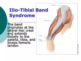
LCL Sprains • Much of the lateral support of the knee is provided by a structure called the iliotibial band • The iliotibial band is often considered to be the true lateral collateral ligament.
LCL Injury Signs & Symptoms • Pain • Lateral knee swelling • Ecchymosis • Point tenderness over the fibular head • Varus instability with second- and third-degree injuries • Pain when the ligament is put on tension during full knee flexion, extension, and varus stress • Limited range of motion
Cruciate Ligaments • The cruciate ligaments are relatively short but strong, rounded bands that cross each other, forming an X within the joint capsule • They are located between the articular surfaces of the tibia and femur and are named according to their tibial attachments. • Their primary function is to provide stability of the knee in an anterior and posterior movement. • They also function to stabilize the knees from rotational stresses, excessive hyperextension.
Cruciate Ligaments • The anterior cruciate ligament (ACL) attaches to the anterior part of the tibia, then crosses upward and backwards to attach on the posterior aspect of the femur. • The posterior cruciate ligament (PCL) attaches posteriorly on the tibia and lateral meniscus, then crosses upward, forward, and inward to a fan-shaped line of attachment on the anterior aspect of the femur.
ACL Sprains • The anterior cruciate ligament an be injured in a number of ways. • Twisting motion during weight bearing • The ACL is often injured along with the medial collateral ligament as a result of a valgus stress to the knee. • Forced hyperextension or a direct blow to the back of the tibia that drives the tibia forward will also cause damage.
ACL Signs & Symptoms • Immediate pain • Unwillingness to move the knee (guarding) • The athlete may also hear a "pop" at the time of the injury. • Joint effusion • Lack of ROM usually results within a 24 hour period • It is important to have the knee examined shortly after the injury.
PCL Sprains • The posterior cruciate may be injured by a direct force against the tibia, which drives it backward in relation to the femur. (90 degrees) • This ligament may also be injured, in conjunction with other supporting structures, from excessive hyperextension, hyperflexion, or abduction forces.
PCL Signs & Symptoms • Pain • Joint effusion • Limited range of motion into full flexion and extension. • With a complete rupture there may be an audible "pop.“ • Athletes who have good quadriceps and hamstring muscle strength may not complain of a feeling of instability with weight bearing, so this type of injury is sometimes may be missed.
Meniscus Injuries • Medial meniscus is damaged more often than lateral meniscus • Medial meniscus attaches directly to medial capsule and MCL • Valgus force can damage meniscus • Rotational force in full flexion or extension- most common way of injuring • Squatting forcefully • Tears in the middle do not usually heal on their own • Lateral tears typically heal with time and rest
Meniscus Injury Signs & Symptoms • Severe Pain • Immediate swelling • Loss of ROM • Locked knee with inability to flex or extend • Pain locally at injured site
Avulsion- indirect fx Comminuted- direct impact Pain Localized edema PRICE X-ray Patellar Fracture
Patellar Subluxations & Dislocations • Athletes predisposed for this problem: • Externally rotated hips • Shallow femoral groves • Flattened lateral femoral grove • Flat patella • Pronated feet • Externally pointing patellas • Weak VMO
Dislocated Patella S & S • Obvious deformity • Pain • Loss of function • Swelling • Ice • Reduce & rehab muscles
Chondromalacia • Softening and deterioration of the articular cartilage on the posterior of the patella • Chronic pain • 3 Stages: • Swelling & softening of Art. Cart. • Cracking of softened cartilage • Surface deformation of Art. Cart.
Chondromalacia • Produced by abnormal patellar tracking • Causes for abnormal patellar tracking: • “knock” kneed (Genu Valgum) • Foot pronation • Externally rotated hips • Shallow femoral groove • Abnormal contour of patella • Laxity of the quadriceps tendon