Download
1 / 44
490 likes | 915 Views
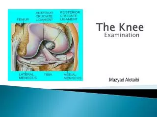
Knee (Tibiofemoral) Joint and Foot. By: Chandie , Christina, Ed & Sharon. Right Tibia and Fibula. Knee Ligaments. Knee Ligaments. Knee Ligaments. Bursae. Suprapatellar bursa Prepatellar bursa Deep infrapatellar bursa Subcutaneous infrapatellar bursa. Bursae.

E N D
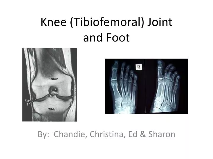
Knee (Tibiofemoral) Jointand Foot By: Chandie, Christina, Ed & Sharon
Bursae • Suprapatellar bursa • Prepatellar bursa • Deep infrapatellar bursa • Subcutaneous infrapatellar bursa
Lateral and Medial Meniscus • Medial meniscus is more “c”-shaped and larger • Lateral meniscus is more circular and smaller • Purpose • Act as cushions • Conforms to the shape of the articulating surfaces as the femur changes position • Provides lateral stability to the knee joint
Knee Muscles - Quadriceps • Rectus femoris • Origin: Anterior inferior iliac spine • Insertion: Tibial tuberosity • Action: Hip flexion, knee extension • Innervation: Femoral nerve • Vascular Supply: Lateral circumflex femoral artery
Knee Muscles - Quadriceps • Vastus lateralis • Origin: Linea aspera • Insertion: Tibial tuberosity via patellar tendon • Action: Knee extension • Innervation: Femoral nerve • Vascular Supply: Lateral circumflex femoral artery
Knee Muscles – Quadriceps • Vastus intermedialis • Origin: Anterior femur • Insertion: Tibial tuberosity via patellar tendon • Action: Knee extension • Innervation: Femoral nerve • Vascular Supply: Lateral circumflex femoral artery
Knee Muscles - Quadriceps • Vastus medialis • Origin: Linea aspera • Insertion: Tibial tuberosity via patellar tendon • Action: Knee extension • Innervation: Femoral nerve • Vascular Supply: Lateral circumflex femoral artery
Knee Muscles - Hamstrings • Biceps femoris • Origin: Long Head- ischial tuberosity; Short Head-Lateral lip of linea aspera • Insertion: Fibular head • Action: Long head- extend hip and flex knee Short head- flex knee • Innervation: Long head-sciatic nerve; Short head-common peroneal nerve • Vascular Supply: Inferior gluteal artery
Knee Muscles - Hamstrings • Semimembranosus • Origin: Ischial tuberosity • Insertion: Posterior surface of medial condyle of tibia • Action: Extend hip and flex knee • Innervation: Sciatic nerve • Vascular Supply: Inferior gluteal artery
Knee Muscles – Hamstrings • Semitendinosus • Origin: Ischial tuberosity • Insertion: Anteromedial surface of proximal tibia • Action: Extend hip and flex knee • Innervation: Sciatic nerve • Vascular Supply: Deep femoral artery
Knee Muscles • Popliteus • Origin: Lateral condyle of femur • Insertion: Posteriorly on medial condyle of tibia • Action: Initiates knee flexion • Innervation: Tibial nerve • Vascular Supply: Popliteal artery
Clinical Concerns: Torn ACL • Purpose of ACL • Prevents anterior translation of the tibia (the tibia moving forward on the femur) • Help maintain alignment of femoral and tibial condyles • Tears can occur due to hyperextension of the knee or excessive inward rotation • Can be due to outside force or non-contact injury • Hear a pop when ACL tears – not all cases • A tear in one of the meniscus is common with ACL tears
Diagnosis and Treatment of ACL Tears • Diagnosis of ACL tears • MRI (magnetic resonance imaging) • X-rays, manual stress tests • Surgical Treatment • Arthroscopic ACL reconstruction • Typically patellar tendon or hamstring grafts • Immobilization brace
Post-Surgery Treatment • First 2 weeks Post-Op • Non-weight bearing • Minimize swelling and regain ROM • Quad sets, straight leg raise, heel slides, knee extensions, CPM machine • 2-6 weeks Post-Op • ROM: continue knee extension and start increasing knee flexion • Exercises: Stationary bike, weight bearing exercises • After 6 weeks Post-Op • Increase strength • No longer need immobilization brace
Post-Surgery Treatment • Conservative and Accelerated rehab protocols • Weight-bearing, ROM, strengthening, agility and brace use vary between the two methods • Custom ACL braces available for physically active or at-risk patients
Tarsal Bones BONES OF THE FOOT
Ligaments: Lower Leg and Foot
Ankle and Foot Muscles • Gastrocnemius • Origin: Medial & lateral condyles of femur • Insertion: Posterior calcaneus • Action: Knee flexion, ankle plantar flexion • Innervation: Tibial nerve • Vascular supply: Popliteal artery
Ankle and Foot Muscles • Soleus • Origin: Posterior tibia and fibula • Insertion: Posterior calcaneus • Action: Ankle plantarflexion • Innervation: Tibial nerve • Vascular supply: Posterior tibial artery
Ankle and Foot Muscles • Extensor digitorumlongus • Origin: Fibula, interosseous membrane, tibia • Insertion: Distal phalanx of four lesser toes • Action: Extends four lesser toes, assists in ankle dorsiflexion • Innervation: Deep peroneal nerve • Vascular supply: Anterior tibial artery
Ankle and Foot Muscles • Extensor hallucis longus • Origin: Fibula and interosseous membrane • Insertion: Distal phalanx of great toe • Action: Extends first toe; assists in ankle inversion and dorsiflexion • Innervation: Deep peroneal nerve • Vascular supply: Anterior tibial artery
Ankle and Foot Muscles • Plantaris • Origin: Posterior lateral condyle of femur • Insertion: Posterior calcaneus • Action: Very weak assist in knee flexion; ankle plantar flexion • Innervation: Tibial nerve • Vascular Supply: Popliteal artery
Ankle and Foot Muscles • Tibialis anterior • Origin: Lateral tibia and interosseous membrane • Insertion: First cuneiform and metatarsal • Action: Ankle inversion and dorsiflexion • Innervation: Deep peroneal nerve • Vascular Supply: Anterior tibial artery
Ankle and Foot Muscles • Tibialis posterior • Origin: Interosseous membrane, adjacent tibia and fibula • Insertion: Navicular and most tarsals and metatarsals • Action: Ankle inversion; assists plantar flexion • Innervation: Tibial nerve • Vascular Supply: Fibular artery
Ankle and Foot Muscles • Flexor hallucis longus • Origin: Posterior fibula and interosseous membrane • Insertion: Distal phalanx of the great toe • Action: Flexes great toe; assists in inversion and plantar flexion of the ankle • Innervation: Tibial nerve • Vascular Supply: Fibular artery
Ankle and Foot Muscles • Flexor digitorum longus • Origin: Posterior tibia • Insertion: Distal phalanx of four lesser toes • Action: Flexes the four lesser toes; assists ankle inversion and plantar flexion • Innervation: Tibial nerve • Vascular Supply: Posterior tibial artery
Ankle and Foot Muscles • Tibialis posterior • Flexor digitorum longus • Flexor hallucis longus • “Tom, Dick & Harry
Ankle and Foot Muscles • Peroneus longus • Origin: Lateral proximal fibula and interosseous membrane • Insertion: Plantar surface of first cuneiform and metatarsal • Action: Ankle eversion; assists ankle plantar flexion • Innervation: Superficial peroneal nerve • Vascular Supply: Fibular artery
Ankle and Foot Muscles • Peroneus brevis • Origin: Lateral distal fibula • Insertion: Base of the fifth metatarsal • Action: Ankle eversion; assists plantar flexion • Innervation: Superficial peroneal nerve • Vascular Supply: Fibular artery
Ankle and Foot Muscles • Peroneus tertius • Origin: Distal medial fibula • Insertion: Base of the fifth metatarsal • Action: Assists somewhat in ankle eversion and dorsiflexion • Innervation: Deep peroneal nerve • Vascular Supply: Anterior tibial artery
Clinical Concerns: Plantar Fasciitis • Plantar fascia • fibrous band that runs from the calcaneus to the base of the toes • Plantar Fasciitis • Inflammation of the plantar fascia • Causes heel pain and can make walking difficult • Risk Factors: • Foot arch problems (flat feet and high arches) • Running • Obesity • Tight Achilles tendon
Plantar Fasciitis • Signs and Symptoms • Sharp pain inside portion of heel • Heel pain that is worse first few steps after awakening, climbing stairs, after long periods of standing • Pain after exercise but not usually during • Mild swelling in heel
Plantar Fasciitis Treatment • Apply ice – ice pack or ice massage • Arch supports or orthotics • Night splints • Stretches for plantar fascia and Achilles tendon • Strengthening for lower leg muscles