Download
1 / 50
560 likes | 1.56k Views
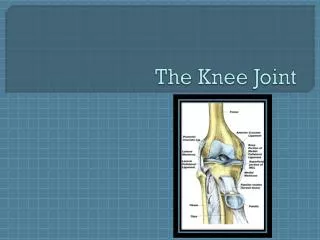
THE KNEE JOINT. Muscles That Act On The Knee. Muscles of the Knee Joint. Hamstrings All - flexion Quadriceps All - extension Unclassified Sartorius - flexion and internal rotation Gracilis - flexion and internal rotation Popliteus - flexion (unlocking) and internal rotation

E N D
THE KNEE JOINT Muscles That Act On The Knee
Muscles of the Knee Joint • Hamstrings • All - flexion • Quadriceps • All - extension • Unclassified • Sartorius - flexion and internal rotation • Gracilis - flexion and internal rotation • Popliteus - flexion (unlocking) and internal rotation • Gastrocnemius - flexion
Anterior Muscles • Quadriceps • knee extension
Rectus femoris • Two joint muscle; most superficial • Origin: anterior-inferior iliac spine of the ilium • Insertion: top of the patella and patellar ligament to the tibial tuberosity • Actions: • Extension of the knee
Vastus lateralis • Origin: Lateral lip of linea aspera. • Insertion: Outer half of the upper border of the patella and the patellar ligament and the anterior tuberoscity of the tibia • Action: • Extension of the knee
Vastus intermedius • Origin: Upper 2/3 of the anterior surface of the femur • Insertion: Upper border of the patella and the patellar ligament • Action: • Extension of the knee
Vastus medialis • Origin: Medial lip of the linea aspera and the internal condyloid ridge • Insertion: Inner half of the upper border of the patella nd patellar ligament • Action: • Extension of the knee
VL RF VM VL RF VM
1 3 2 Hamstrings Quadriceps • Vastus Lateralis • Vastus Intermedius • Rectus Femoris • Vastus Medialis
QUADRICEPS MUSCLES • Vertical jumping • Decelerating or eccentric contraction during landing or changing directions • As a group, they are typically 25-30% stronger than the hamstrings
Hamstrings • knee flexion
Biceps femoris • Lateral side • Origin: • 1.) Long head - ischial tuberosity; • 2.) Short head - lower half of the linea aspera • Insertion: Head of the fibula • Action: • Flexion of knee
Semitendinosus • Medial side; superficial • Origin: Ischial tuberosity • Insertion: Anterior medial surface of the tibia • Action: • Flexion of the knee
Semimembranosus • Medial side, deeper than semitendonosus • Origin: Ischial tuberosity • Insertion: Anterior medial surface of the tibia • Action: • Flexion of the knee
HAMSTRING MUSCLES • More “knee flexors” than “hip extenders” • Acceleration muscles during running • Tight hamstring muscles can contribute to lower back, hip and knee problems
Gracilis • O: Pubis crest • I: Anterior medial surface of the tibia • Actions: • flexion at the knee
Sartorius • Posterior at the knee joint • Origin: Anterior-superior spine of the ilium • Insertion: Anterior medial surface of the tibia • Action: • Flexion at the knee • From Late Latin sartor, tailor (from its producing the cross-legged position of a tailor at work)
Popliteus • Origin: Lateral condyle of the femur • Insertion: Proximal third of posterior aspect of the tibia • Action: • Flexion of the knee • It "unlocks" the knee. • Only true knee flexor
Gastrocnemius • Origin: posterior surface of the medial and lateral femoral condyles • Insertion: the calcaneus through the Achilles tendon • Actions: • Flexion of the knee Posterior
Review • Superficial Leg Muscles
Name the muscle and its action(s) Sartorius Action: Flexion at the knee
Q Angle Valgus Varus The deviation between the line of pull of the rectus femoris and the patellar ligament. Stress to lateral side of the knee Stress to the medial side of the knee Terms
Semimembranosus Name the muscle and its action(s) • Action: • Flexion of the knee
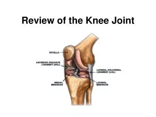
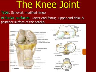
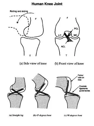
Anterior cruciate ligament • Lateral collateral ligament • Lateral meniscus • Posterior cruciate ligament • Medial collateral ligament • Medial meniscus 1. 2. 3. 4. 5. 6.
Semitendinosus Name the muscle and its action(s) • Action: • Flexion of the knee
1 = PCL 2 = Lateral meniscus 3 = ACL 4 = Medial meniscus 1. 4. 2. 3
Name the colored muscles 2 = Vastus lateralis 1 = Rectus femoris 4 = Vastus medialis
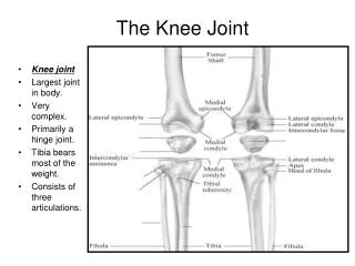
? = ? = ? = Lateral condyle Medial condyle Intercondylar fossa Femur
Name the muscle and its action(s) Gracilis Action - flexion at the knee
? =? = ? = Medial condyle Lateral condyle Medial Malleolus Tibia
2. 3. 1. • = Vastus lateralis • = Rectus femoris • = Vastus medialis
VL RF VM VL RF VM
Rectus femoris Name the muscle and its action(s) • Actions: • Extension of the knee
1. 2. 3. 3. 4. 1. = Anterior cruciate ligament 2. = Lateral collateral 3. = Lateral meniscus 3. = Medial collateral 4. = Medial mieniscus
Name the muscles. 1 = Vastus lateralis 2 = Vastus intermedius 3 = Vastus medialis 1. 2. 3.
Fibula ? = ? = Head Lateral malleolus
Name the muscles • Biceps femoris • Biceps femoris • Semitendonosis • Semimembranosis
Biceps femoris Name the muscle and its action(s) • Action: • Flexion of knee
Medial condyle Lateral condyle Medial Malleolus Tibia ? =?= ? =
Popliteus Name the muscle and its action(s) • Action: • Flexion of the knee
6 = ? 7 = ? 8 = ? Lateral condyle Intercondylar fossa Medial condyle Femur
Name the muscle and its action(s) Sartorius Action: Flexion at the knee
3. 1. 4. 2. 1 = Medial meniscus 2 = PCL 3 = ACL 4 = Lateral meniscus