Download
1 / 62
650 likes | 1.19k Views
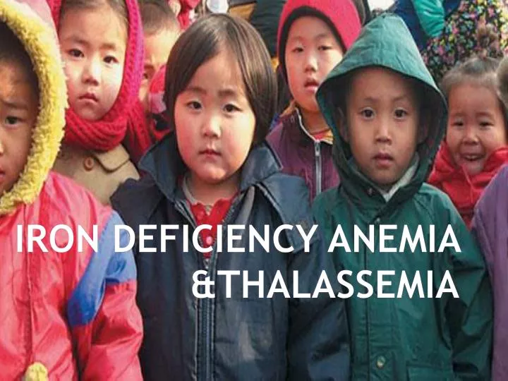
IRON DEFICIENCY ANEMIA &THALASSEMIA. B Major Thalassemia. Anemia. It is a reduction of the red cell volume or hemoglobin concentration below -2SD for age, sex. Normal Range,Hb. Birth:16/6 gr/dl 2 Mo:11/5 3-6 Mo 11/5 6-24 M :12 2-6 Y:12/5 6-12 Y:13/5 12-18 Y: F =12-14 M=14-16.

E N D
Anemia It is a reduction of the red cell volume or hemoglobin concentration below -2SD for age, sex.
Normal Range,Hb • Birth:16/6 gr/dl • 2 Mo:11/5 • 3-6 Mo 11/5 • 6-24 M :12 • 2-6 Y:12/5 • 6-12 Y:13/5 • 12-18 Y: F =12-14 M=14-16
Normal Range,MCV • Birth:108FL • 2 Mo:96 • 3-6 Mo:91 • 6-24 M :78 • 2-6 Y:81 • 6-12 Y:86 • 12-18 Y: F =90 M=88
Iron deficiency Folate deficiency Hemoglobinopathies Vitamin B12 deficiency Anemia HIV infection Malaria Infectious/inflammatory disorders Helminth infection
Red blood cells specialisations biconcave shape no nucleus extra space inside contain haemoglobin the oxygen carrying molecule increases the surface area so more oxygen can be carried
Iron deficiency is a major health problem worldwide and especially in developing countries. • Iron-deficiency is the most prevalent nutritional deficiency worldwide • Iron deficiency is the most common single cause of anemia worldwide
Review Of Articles 1 • Prevalence of iron deficiency anemia in 6mo-5 years old children in Fars , southern IRAN • Kadivar MR & Collegues.Med Sci Monit,2003;9(2);CR 100-104 • 541 patients: • 110 p(%19.7): Serum Ferritin level < 12ng/ml • 101 P(%18.7): low serum Hb • Developing Countries: IDA%25-%35 • Industerialized Country: IDA %5-%8 • Iron supplements by Health care centers , Free of charge
Ironstatus The concentration of Iron in: Infant: 75-80 mg/kg(BW) 50mg/kg: Hb Mass 25mg/kg: Storage Iron 5mg/kg: Myoglobin & tissue Iron
Ironstatus The concentration of Iron in: Adult: 40-50 mg/kg(BW) 30mg/kg: Hb Mass 6-7mg/kg: Myoglobin, Heme enzymes & non heme enzymes * 6-7mg/kg (F) storage Iron * 10-12 mg/kg (M) < 0/5%: Transport Iron
IronMetabolism • Cellular sequestration & Metabolism of Iron is mediated by 3 proteins: • Transferrin • Transferrin receptor • Ferritin
Ferritin Ferritin is the major storage protein with 24 subunit: * Light chain (L), 19 kD * Heavy chain (H) 21 kD H gene locus: ch 11: Heart, Iron – Metabolism L gene locus: ch 19: Liver & spleen- Iron storage function Ferritin is found in virtually all cells especially: Erythroied precursors Macrophages Hepatocytes F.molecule: 4500 Iron atoms Half life: 60 hour Catabolism of, F: Reutilization of Iron core Hemosiderin conversion
Body Iron Distribution and Storage Duodenum Dietary iron (average, 1 - 2 mg Utilization Utilization per day) Plasma (TIBC) transferrin (3 mg) Bone Muscle marrow (myoglobin) (300 mg) Circulating (300 mg) erythrocytes Storage (hemoglobin) iron (Ferritin) (1,800 mg) Sloughed mucosal cells Desquamation/Menstruation Other blood loss (average, 1 - 2 mg per day) Reticuloendothelial Liver macrophages (1,000 mg) Iron loss (600 mg)
Iron balance • Iron balance is physiologically regulated by controlling Iron absorption. • The availability of dietry Iron for absorption is dependent to: • The amount of Iron • Form of Iron • Composition of the diet • GI factors
CBC • Anemia Hb, HCT • leukocytosis • leukopenia • Abnormal cells WBC -Thrombocytosis -Thrombocytopenia Platelet
peripheral Blood,CBC • R BC • Hb (is not specific) • MCV • MCH • Reticulocyte • Peripheral blood smear, Morphology
Indirect: 1- plasma ferritin: (the most useful) in the absence of: * Tissue necrosis * Inflammation * Neoplasm * liver disorder * turn over of RBC
Prussian Blue Stainof Bone Marrow Iron Present No Iron Present
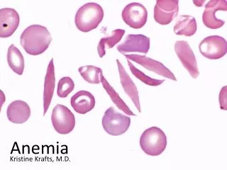
Iron deficiency anemia Iron deficiency anemia is the most common cause of anemia. Growth & diet are almost always contributing factors in childhood
Red Blood Cells Etiology / IDA • Blood Loss • Gastrointestinal Tract: • Milk -induced Enthropathy • Peptic ulcer • Inflamatory Bowel Diseaes • Meckel Diverticuculm &Polips • Drugs: Salicylates • Hookworm Infestation • Pulmonary Hemosiderosis • Iatrogenic • Menstural Blood Loss • Urinary Blood Loss(rare)
Etiology/IDA • Increased Physiologic Requirement -Pregnancy -Infancy -Adolescence • Malabsorption - Inflamatory Bowel Diseaes -Tropical Sprue • Gastrectomy • Pica • Dietary inadequacy: Iron Poor Diet • Combinations of above
Clinical manifestations • *Hematologic • * Non Hematologic • Pallor • Weakness, fatigue, Irritability • Anorexia • Pica • Blue sclera • Koilonychias (spoon- shaped nails) • Glossitis • Angular stomatatis • Post cricoid esophageal web (plummer winson syndrome) • Impair of intellectual & learning • Impaired of immunity • Slightly enlarged spleen • Cardiopulmonary failure & death.
Laboratory test: 1-Serum Ferritin: < 10-12 ug/l 2- Serum Iron( Decrease) 3-Total iron binding capacity TIBC 4- peripheral blood : RBC, Hb- HCT↓ MCV, MCH ↓ (RDW(Red blood cell distributaion width ) Reticulocyte , Mild 4- Serum Soluble Transferrin Receptor 5-FEP 6- BMA & BM Biopsy (Prussian Blue Staining)
Reticulocyte count Normal = 0.2-2 % Corrected reticulocyte = Pt HCT X Reti. Normal HCT
Anemia Hb, HCT • leukocytosis • leukopenia • Abnormal cells WBC -Thrombocytosis -Thrombocytopenia Platelet CBC
Differential Diagnosis of I.D.Anemia 1- . Thalassemia minor 2- . Thalassemia major 3- Chronic disorders 4- lead poisining 5- . Thalassemia
B Thalassemia trait (Heterozygous) • Expression of one gene is impaired by mutation where as the other gene is normal. • Slight ineffective erythropoiesis & modestly decrease of RBC survival • Mild erythrocytosis • Marked microcytosis • Peripheral Blood: microcytosis, hypochromia & targeting
Differntial Diagnosis B Thalassemia trait / Iron Deficiency Anemia B. Th. Trait: • Increase of RBC- Mild Erythrocytosis, • Marked microcytosis • IDA : RBC count decreased, MCV is rarely as low as B. Th .Trait • RDW ( Red Cell Disrtribution Width by Automated cell counter) : Increased in IDA • Mentzer Index( MCV/RBC ): • B .Th .Trait <13 • IDA > 13
CBCB. TH .Trait &I. D.A WBC=10000/mm3 WBC=6000/mm3 RBC=6/000/000/mm3 RBC=3/200/000/mm3 Hb=10 gr/d Hb=7gr/dl HCT=%30 HCT=%21 MCV=60 FL MCV=74FL MCH=23 pg MCH=25Pg Platelet=180000/mm3 Platelet=600000/mm3
a Hb A HbA2 b g Hb F d b thalassemia
B Thalassemia trait (Heterozygous) • Hb Electrophorasis: High A2 Hb (3.5- 8%) High A2 & High F Hb(5%-20%) Low A2 Hb (Hb F 5%-15%, Thalassemia) Normal A2 Hb
IRON DEFICIENCY versus ACD Serum Iron Transferrin Ferritin Iron Deficiency ACD
Major Thalassemia /Cooley Anemia • Compound heterozygous state for two different globin gene mutations • Homozygous state for the same mutation. • Age of diagnosis: 6-12 months • 60%: first year • 29%: second year • 9%: later
Clinical manifestations • Pallor • Failure to thrive • Irritability • Icterus • Hepatosplenomegaly • Skeletal changes • Prone to infection