Download
1 / 19
190 likes | 285 Views
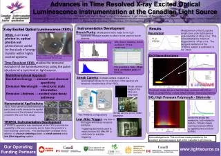
Towards X-ray excited optical microscopy (XEOM) for cultural heritage, spectroelectrochemistry, and wider applications. Mark Dowsett 1 , Annemie Adriaens 2 , Gareth Jones 1 and Alice Elia 2. 1 Analytical Science Projects Group, University of Warwick

E N D
Towards X-ray excited optical microscopy (XEOM) for cultural heritage, spectroelectrochemistry, and wider applications Mark Dowsett1, Annemie Adriaens2, Gareth Jones1 and Alice Elia2 1Analytical Science Projects Group, University of Warwick 2Electrochemistry and Surface Analysis Group, Ghent University Thanks to : Paul Thompson, Simon Brown (XMaS, ESRF) Sergey Nikitenko (DUBBLE, ESRF), Nigel Poolton (Formerly SRS, Daresbury) Analytical Science Projects
Goal: Develop XEOL microscope coupled to an environmental cell for synchrotron applications Real time process monitoring in controlled electrochemical and gaseous ambients – corrosion and protection studies Ultimate goal: Develop a portable version for direct chemical imaging in museums etc. Microscopy of the chemical state rather than just elemental composition (i.e. A step beyond portable XRF)
Why XEOL? • Based on transoptical emission (200-1000 nm) caused by keV X-ray irradiation - phosphorescence, fluorescence • Electronic processes responsible for XANES and EXAFS impose similar structure on the light emission • Extra band specific-features due to excitation of chromophores by LE electron scattering • Spectra are (at least) two dimensional – X-ray energy and emitted optical wavelength • Technique has a high surface specificity – sees thin layers on surfaces invisible to conventional XAS • Basis of a chemically specific optical microscopy - image formation using broadband light optics
Shutter Web cam (1 of 2) Stepper 1 Filter housing X-ray port 1 Ref. electrode Broadband PM Optical bench Illumination Stepper 2 Optics X-ray port 2 eCell X-ray detector Proof of concept - ODXAS 1
Filter sample Silica condenser Silica objective window piston
Copper – XAS and XEOL-XAS XAS (DUBBLE) XAS (DUBBLE) XAS (DUBBLE) XEOL (XMaS) XEOL (XMaS) Parallel XAS (XMaS)
Cuprite (Cu20) – XAS and XEOL-XAS Comparison Dowsett, Adriaens, Jones, Fiddy, Nikitenko, Anal. Chem. 80 (2008) 8717-8724
Materials of construction ... and the rest ... ... e.g. The eCell window eCell body X-ray shielding Optical column Substrate (for Powders etc.) (13 keV X-rays)
Other edges (so far) Pb (LIII) Zn (K) Pb (LIII) 1.8 eV Clean metal Lead decanoate
Colour filters Nantokite (CuCl) on copper Atacamite (Cu2(OH)3Cl) on copper
Identifying mixtures with filters Thick layer on copper “Fine dusting” on acetal Through green filter
Potential imaging modes Time to a mean 3% precision per pixel in broadband image < 1000 s for 2048 x 2048 pixels • Filtered images • Dispersed images • Near edge image spectra • Edge correlated images (form image on correlation with specific oxidation state etc. • EXAFS image spectra – given time
Next steps Image Broadband CCD 2, 2048 x 2048 Filtered (imaging) or broadband (spectroscopy) Filters (imaging mode) Focusing condenser Objectiveoptics Broadband optical emission X-rays Sample Schematic diagram of XEOM 1 - Imaging
Next steps Projection optics Grating Spectrum/Image spectrum Broadband CCD 1, 500 x 2048 Focusing condenser Objectiveoptics Broadband optical emission X-rays Sample Schematic diagram of XEOM 1 - Spectroscopy
Summary and Conclusions • Silica lens – based microscope –> mid to end 2010 • Mirror-based device -> 2011-2012 • Portable device ? • XEOL provides multispectral information including XANES and EXAFS from heritage metals and corrosion products • Optical devices with constructed with broadband optics will provide microscopy with (light) wavelength limited resolution • Suitable for beam lines with large (millimetres ) footprint • XEOM has potential applications in • Measurements in controlled environments • Metal corrosion research • Geosciences • Semiconductor research • Organo-metallics • ...