Download

1 / 19
190 likes | 382 Views
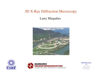
3D X-Ray Diffraction Microscopy. Larry Margulies. 200 µm. Metal Structures. Defor- mation. Heat. 200 µm. 5 µm. Challenges: - Multiple lengthscales - Heterogeneities - Predicting the dynamics. Traditional Microscopy is 2D. 4D (space + time). 3DXRD Vision.

E N D
3D X-Ray Diffraction Microscopy Larry Margulies
200 µm Metal Structures Defor- mation Heat 200 µm 5 µm Challenges: - Multiple lengthscales - Heterogeneities - Predicting the dynamics Traditional Microscopy is 2D 4D (space + time)
3DXRD Vision • 3D characterization of individual grains within bulk polycrystals • Volume • Crystallographic orientation (intragranular ODF) • Grain boundary morphology (3D mapping) • Elastic strain tensor • Structural refinement • Statistics over 100-1000 structural units • In-situ annealing and deformation studies(4D)
3DXRD set-up Area detector Detector I L = 5-10 mm Position and Orientation Detector II L = 40 cm Orientation and Strain Acq. time: 1-10 sec 10-100 msec
Two step process 1. Indexing: GRAINSPOTTER IMAGED11 2. Reconstruction GRAINSWEEPER Up to 1000’s grains: CMS position volume: 1-10% average orientation: 0.2 deg average elastic strain: De =10-4
Growth curves: New Avrami-type model Statistics 244 grains log(-ln(1-VV)) 0 1 2 log (time) Growth curves Farfield detector only:
Phase Transformations in Carbon Steel Pearlite – Ferrite – Austenite g q g g g a a q a a a b Growth curves N dN/dt Activation energy off by 100!
Grain rotation Grain rotation for 95 grains in Al, 100 mm grains, 5 mm thick Tensile strain: 6 %
Multicrystal Third route in Crystallography Single Crystal Powder X-ray data:
Applications to: • Pharmacy • Photochemistry • Protein Crystallography Third route in Crystallography Validation: Cu(C2O2H3)2.H2O. 70 grains of size < 1 micron Cell ~1400 Å3 (C2/c) Result: Single crystal quality refinement!
Grain Mapping Area detector Detector I L = 5-10 mm Position and Orientation Detector II L = 40 cm Orientation and Strain
Video of growth of an internal grain Recrystallization of 42% deformed pure Al during annealing at ~200 C.
Grain growth Sample: Al(0.1% Mg) Initial 800 min anneal at 450 C 491 grains 49 grains
CCD YAG 150µm YAG:Ce 25µm Sample Present detector • Resolution: ~3µm • Efficiency: ~1% • Long tails in PSF
Structured Scintillator Principle: Electrochemical etch: @ KTH, Sweden
LuAG 25µm SS 4µm pitch Tomographic images of Al with W particles taken with a conventional LuAG 25µm thick screen and a structured scintillator with a 4µm pitch
Future of 3DXRD faster and smaller • 3D high resolution detector • mapping of deformed microstructures • “box beam” mapping • Faster throughput for in-situ mapping • 100 nm resolution 2D(3D) detector • R&D is needed for a solid state device
Acknowledgments Risø:H.F. Poulsen, C. Gundlach, D. Juul Jensen, E. Knudsen, E.M. Lauridsen,W. Pantleon, S. Schmidt, H.O. Sørensen, G. Winther ESRF, ID11:A. Goetz, Å. Kvick, G. Vaughan, J. Wright
3DXRD Instrumentation Optics (brown): WB: White beam LC: Bent Laue crystal WBS: White beam stop ML: Bent multi layer MB: 2 dimensionally micro focussed monochromatic beam BS: Monochromatic beam stop Sample environment (yellow): I: Cryostat II: Furnace III: 24kN Stress rig Detectors / slits (purple): 1: Large area detector 2: Conical slit system 3: High resolution area detector 4: Optional detector system 5: Small area detector