Download
1 / 23
230 likes | 238 Views
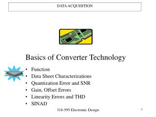
ACRIN-6684 - MRS data acquisition and Raw Data Handling Instructions – For Siemens Data. In vivo MR Spectroscopy. Representative MRS of a normal human brain @3T. NAA. Cho. Cr. Lipids, macromolecules. Glu/ Gln. MI. Proton MRS is able to detect the following metabolites:

E N D
ACRIN-6684 - MRS data acquisition and Raw Data Handling Instructions –For Siemens Data
In vivo MR Spectroscopy Representative MRS of a normal human brain @3T NAA Cho Cr Lipids, macromolecules Glu/ Gln MI
Proton MRS is able to detect the following metabolites: • N-Acetyl Aspartate (NAA) at 2 ppm: Marker of neuronal density and viability • Creatine (Cr) at 3 ppm: Energy metabolism, generation of ATP • Choline (Cho) at 3.2 ppm: Pathological alterations in membrane turnover, increased in tumors • Lipids (Lip) between 0.8 – 1.5 ppm: Breakdown of tissue, elevated in brain tumors - lipids indicate necrosis
Cho NAA Cr Lac • Lactate (Lac) at 1.3 ppm, inverted at 144ms: produced by an anaerobic metabolism, found in tumor containing zones of necrosis
The Sequence 3D chemical shift imaging using a Point-resolved spectroscopy (PRESS) excitation pulse sequence. 3D Volumetric Spectroscopy preferred 2D CSI Spectroscopy is acceptable
Optimal Voxel Placement The ROI will be placed at the center of the enhancing tumor covering the lesion and the normal brain as much as possible but excluding the subcutaneous fat and sinuses.
Suboptimal Voxel Placement Proximity to sinuses can result in signal broadening and susceptibility artifacts Proximity to scull can result in contaminating lipid signal
3D MRSI Parameters • TE 144 ms, TR 1140 ms, • FOV > 160 mm2, • Phase encoding arrays 12 x 12 x 8 • Numbers of Acquisitions: 1 • Spatial zero-filling to 16 x 16 x 8 phase encoding arrays will result in an individual voxel size of 1 x 1 x 1 cm3. • Approximate imaging time: ~6 min utilizing elliptical k-space sampling k-space sampling • Manual shimming is recommended before the acquisition to obtain the best magnetic field homogeneity.
Shimming “Shimming” = adjusting the magnetic field to make it more homogeneous 1.5T: Signal line width or full width at half maximum (FWHM): <15 Hz for 3D MRSI 3 T: FWHM < 25 Hz for 3D MRSI NAA Cho&Cr • Better signal separation, thus better quantification of metabolites • Better water suppression • Suboptimal shimming
Manual Shimming on Siemens Hit the “Show” tab Then “Invalidate All” “Adjust All” Wait a couple of minutes until it says “Adjustments finished”
Hit the “Interactive Shim” tab Then hit “Measure” (1) Numbers will begin to scroll on the above white box (2) You can alter the shim by changing Z, then Y, then X. Do this slowly, only a couple of increments at a time, then start with z again … Stop (3) and Apply (4) 2. 1. 3. 4.
Saturation Bands • Click SAT and place up to 10 SAT bands around the voxel to suppress signals from lipid/fat
The spectrum appears in one of the quadrants • Note: if another window appears asking you to select a protocol for the spectrum, just select any of the protocols listed and click OK.
Go to the top of the screen Click on “Patient” Dropdown menu will appear “Save data”
Select first option, Selected results and 3 reference images (shown above) • This will save the spectrum and corresponding images in a DICOM format
In order to use a different slice: Right click on the Image A scroll down window will appear Select 3D CSI Selection
Under 3D CSI Selection: Choose Plane number
Saving the Data in .rda format • Click on Options – export raw data (shown above).
Saving the Data in .rda format • The previous command (Options-export raw data), will open this new window. By default the directory is C:\Temp (leave default) • .Save File name as “ACRINcase#_timepointXweeks” • For example: case15_baseline.rdaor case09_16weeks.rda • Click Export