Download
1 / 34
340 likes | 413 Views
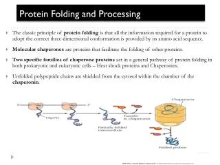
Evidence for Abnormal Protein Processing in AD. Carl W. Cotman Institute for Brain Aging and Dementia UC Irvine. Examples of Abnormal Protein Processing. APP generating β -amyloid Cytoskeletal proteins: hyperphosphorylation and proteolysis.

E N D
Evidence for Abnormal Protein Processing in AD Carl W. Cotman Institute for Brain Aging and Dementia UC Irvine
Examples of Abnormal Protein Processing • APP generating β-amyloid • Cytoskeletal proteins: hyperphosphorylation and proteolysis
Proteins implicated in AD are targets of caspases Amyloid precursor protein (APP) Gervais et al. (1999) Cell97: 395-406 Lu et al. (2003) J Neurochem87: 733-41 Presenilin-1 (PS1) van de Craen et al.(1999) FEBS Lett 445: 149-54 Fluhrer et al. (2004) J Biol Chem279: 1585-93 Tau Canu et al. (1998) J Neurosci18:7061-74 Fasulo et al. (2000) J Neurochem75: 624-33 Gamblin et al. (2003) PNAS100: 10032-7
Caspase activation in the AD brain: usually chronic Stadelmann (1999) Am J Pathol 155:1459-1466 Su et al. (2001) Brain Res 898:350-7 Rohn et al. (2001) Neurobiol Dis 8:1006-16 Rohn et al. (2002) Neurobiol Dis 11:341-54 Su et al. (2002) Acta Neuropathol 104:1-6, 2004 Gastard et al (2003) Ann Neurol 54:393-8 Pompl et al. (2003) Arch Neurol 60: 369-76.
Biology of Tau • Tau is a microtubule associated protein that drives microtubule assembly therby stabiizing the cytoskeleton, • tau also participates in vesicular transport and axonal polarity. • 6 isoforms of Tau exist in the adult human brain all of which are produced by alternative splicing from one gene located on chromosome 17. • These isoforms of tau differ by the inclusion or exclusion of 1 or 2 n-terminal inserts and/or a fourth microtubule binding domain (3R vs 4R). • All 6 isoforms of tau contain a caspase 3 and 7 consensus sequence (DMVD).
Tau is a microtubule associated protein Spillantini et al., (1998) Trends in Neuroscience21: 428-33
Alterations of tau conformation and phoshphorylation in AD Sequence:1.MC1, 2. AT8, 3.PHF-1Caspase clavage?
Tau is cleaved at Asp421 Dtau C-terminus
Caspase-cleavage of tau induces a conformational change recognized by the early-tangle marker MC1
Dtau is hyperphosphorylated in vitro by GSK-3b: PHF-1 positive
Antibodygeneratedis specific for tau cleaved after Asp421 generated by executioner caspases
Dtau is detected in the AD brain AD Control Preadsorption
The α-DTau antibody specifically recognizes caspase-cleaved tau in vivo: does not stain tau-/- mouse brainafter head injury DTau Fodrin-CCP Caspase-3
Caspase-Cleavage of Tau is Correlated with Cognitive Decline r = -0.857 MMSE Cell Number The increased presence of the number of DTau positive cells was inversely correlated with cognitive decline, as determined by MMSE score
Dtau becomes increasingly insoluble with AD progression RAB RIPA
Tau Pathology in Nondemented and Early AD Cases in area CA1 of the hippocampus
Tau Pathology in Nondemented and Early AD Cases in area CA1 of the hippocampus
Dtau and full-length tau are both present within NFTs and dystrophic neurites
What leads to Dtau in AD? Does Aβ drive tau pathology? Ab activates caspases in vitro: Loo et al. (1993) PNAS 90:7951-5 Ivins et al. (1998) Neurobiol Dis 5:365-78 Oxidative stress activates caspases in vitro: Camondola et al. (2000) J Neurochem 74:159-68
Is cleaved tau present in 3xTg-AD mouse and does Dtau co-localize with Ab1-42 ?
Does Dtau co-localizes with MC1 in 3xTg-AD mouse?
Summary • Several proteins get cleaved by caspases and appear to be present in neurons for prolonged periods of time, e.g., fodrin, actin and tau as well as APP • Tau is cleaved by executioner caspases initiated by β-amyloid, oxidative damage • Cleaved tau seeds (nucleates) the assembly of tau into PHF-1 like assemblies and assumes an MC-1 conformation. • Cleaved tau is present in pre-tangle and tangle neurons • Cleaved tau neurons inversely correlate with cognitive function • Chronic abnormal protein processing may be a new mechanism catalyzing AD pathology
Acknowledgements Cotman Lab Dr. Carl Cotman Dr. David Cribbs Dr. Elizabeth Head Christina Tu Dr. Reidun Torp Dr. Paul Adlard Dr. Pat Kesslak LaFerla Lab Dr. Frank LaFerla Salvatore Oddo Boise State U. Dr. Troy Rohn Mass spectroscopy Nemone Muster Laser Light Scattering Dr. Wytze van der Veer
The C-terminal-specific antibody T46 does not recognize Dtau
g-Secretase substrates integral to AD Pathogenesis Substrates APP & APPLPs E-Cadherin Notch 1-4 ERB-4 Nectin-1a CD44 LRP P75 Components of the g-Secretase Complex g-Secretase Complex adapted from Kimberly & Wolfe, 2003
AD pathologic hallmarks: Senile plaques and neurofibrillary tangles