Download
1 / 36
420 likes | 754 Views
8 Protein Synthesis, Processing, and Regulation. Chapt 8 Student learning outcomes Because proteins are the active players in most cell processes Explain general process of Translation of mRNA: indicate similarities, differences prokaryotes, eukaryotes

E N D
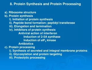
8 Protein Synthesis, Processing, and Regulation • Chapt 8 Student learning outcomes • Because proteins are the active players in most cell processes • Explain general process of Translation of mRNA: indicate similarities, differences prokaryotes, eukaryotes • Explain that protein function requires proper folding and processing, including sorting and transport • Describe several mechanisms of regulation of protein activity • Describe 2 ways for protein degradation: • Ubituitin-proteasome; lysosome
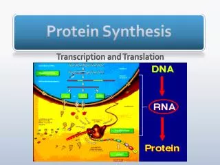
Introduction Translation: synthesis of polypeptide directed by mRNA template on ribosome mRNAs read 5′ to 3′ direction; polypeptide synthesized from NH2 to COOH terminus. Amino acid specified by 3 bases (codon) in mRNA rRNA, tRNA, mRNA roles Translation is first step to form functional protein: polypeptide chain must fold into appropriate conformation often undergoes processing steps. Gene expression is regulated at level of translation Many controls on amounts and activities of proteins: ultimately regulates all aspects of cell behavior.
Translation of mRNA tRNAs (70 - 80 nt) align amino acids with codons on mRNA Cloverleaf Structure; CCA at 3′ terminus, anticodon loop binds to codon (complementary bp) Aminoacyl tRNA synthetases Attach amino acids to specific tRNAs Each enzyme recognizes one amino acid, as well as correct tRNA(s) Costs ATP to attach . Figs. 8.1,2
Translation of mRNA Ribosomes named according to sedimentation rates in ultracentrifugation Ribosomes are abundant in cells lot of protein synthesis; E. coli ~ 20,000; mammalian cells ~ 10 x 106 Fig. 8.4 B, 30S, C, 50S
Translation of mRNA Evidence rRNA does catalysis: Noller et al. (1992): large ribosomal subunit could catalyze formation of peptide bonds even after 90% of ribosomal proteins were removed. High-resolution structure of 50S (Steitz, 2000): Ribosomal proteins absent from site of peptidyl transferase reaction. Ribosomal proteins mostly structural Nobel Prize 2009 ribosome structure Fig. 8.6
Fig 8.7 Prokaryotic and eukaryotic mRNAs • mRNAs contain untranslated regions (UTRs 5’, 3’) • Eukaryotic mRNAs usually one polypeptide chain • Monocistronic • Prokaryotic mRNAs often encode many polypeptides • Polycistronic(e.g., lacoperon). Fig. 8.7*
Translation of mRNA Translation initiates with Met, usually 5’-AUG. Bacterial mRNA AUGs preceded by Shine-Dalgarno sequence - aligns mRNA on ribosome Eukaryotic mRNAs recognized by 7-MeG cap at 5′ terminus. Ribosomes scan downstream until initiation codon. Fig. 8.8
Translation of mRNA Translation: initiation, elongation, and termination. Initiator met-tRNA and mRNA bind small ribosomal subunit. Large ribosomal unit joins, forming functional ribosome. Fig. 8.9*
Many non-ribosomal proteins for initation. • Initiation starts with 30S ribosomal subunit bound to IF1 and IF3. • mRNA, initiator N-formylmethionyl (fMet) tRNA and IF2 (bound to GTP) join • IF1 and IF3 release, 50S subunit binds, • IF2-GDP is released • 5’-CAU of tRNA binding 5’-AUG of mRNA Fig. 8.10 Bacterial initiation
Fig 8.12 Elongation stage of translation • Elongation similar in prokaryotes and eukaryotes • 3 binding sites: P (peptidyl), A (aminoacyl), and E (exit). • Initiator met-tRNA bound at P site. • elongation factor (EF-Tu prokaryotes, eEF1a in eukaryotes) • bound to GTP brings aminoacyltRNA to complex • Translocation – • Ribosome moves 3 nt • Next codon in empty A site. • PeptidyltRNA from A to P, • Uncharged tRNA from P to E • New aa-tRNA binds A site • Uncharged tRNA leaves Fig. 8.12
Fig 8.14 Termination of translation Termination: elongation continues until stop codon (UAA, UAG, or UGA) translocated into A site. Release factors recognize codons, terminate Fig. 8.14 termination
Translation of mRNA Polysomes (polyribosome) : mRNAs translated simultaneously by several ribosomes Once ribosome moved from initiation site, another can bind. begin synthesis Fig. 8.15 polysomes
Translation of mRNA Regulation of translation modulates gene expression: translational repressor proteins noncoding miRNAs, siRNA, RNAi localization of mRNAs Ex. Cis-acting sequence in mRNA binds repressor Translation of ferritin mRNA regulated by repressor proteins. Iron absent, iron regulatory protein (IRP) binds iron response element (IRE) in 5′ UTR, blocks translation Iron present, get translation Fig. 8.16 eukaryote Ferritin regulation
Fig 8.18 Localization of mRNA in Xenopus oocytes • Ex. Localization of mRNAs to specific regions of eggs or embryos important in development: • permits proteins synthesized at appropriate sites. • Proteins binding 3′ UTRs can localize mRNAs to specific • regions of cells. • mRNAs with short poly-A tails are stored in oocytes; • translation activated at fertilization or later • Lengthening poly-A tails allows binding of poly-A binding • protein (PABP), stimulates translation Fig. 8.18 mRNA localization Xenopusoocyte
Translation of mRNA RNA interference (RNAi) short ds RNAs block translation Small interfering RNAs (siRNAs)— ds RNAs, nuclease Dicer. MicroRNAs (miRNAs)— transcribed by RNA pol II, cleaved by nucleases Drosha and Dicer. RNA-induced silencing complex (RISC): siRNAs or miRNAs that pair perfectly induce cleavage of targeted mRNA; most miRNAs form mismatches, repress translation Fig. 8.19 RNAi
Translation of mRNA Modification (phosphorylation) of initiation factors can regulate translation global effects on overall translational activity Ex. Phosphorylation of eIF2, eIF2B by protein kinases blocks exchange of bound GDP for GTP, inhibits initiation. Fig. 8.20
Protein Folding and Processing *2. Protein folding, processing is critical: Polypeptide chains must undergo folding, other modifications, to become functional proteins Information for conformation comes from amino acid sequence. Folding and Processing includes: Chaperone proteins S-S bonds between Cys residues [Peptide bond isomerization (Pro residues)] Proteolytic cleavage (removal of Met, pre-sequences) Glycosylation (addition of sugars) Addition of lipids
Protein Folding and Processing Chaperones: facilitate folding of other proteins. Catalysts - facilitate assembly, are not part of complex. Bind, stabilize unfolded or partially folded polypeptides Protect chain from aberrant folding or aggregation until synthesis of an entire domain is complete Stabilize unfolded polypeptide chains during transport into organelles; later assist refolding Figs. 8.22, 23
Fig 8.24 Sequential actions of chaperones Chaperones found as heat-shockproteins (Hsp) • Expressed in cells subjected to high temperatures. • Stabilize, facilitate refolding of partially denatured proteins Chaperonins -protein subunits in stacked rings (double-chambered structure) • isolates protein from cytosol, other unfolded proteins [Enzymes can be chaperones: protein disulfide isomerase, peptidylprolylyisomerase] Figs. 8.24
Protein Folding and Processing Proteolytic processing - cleavage of polypeptide 1. Removes portions - initiator Met from NH2 terminus. 2. NH2-terminal signal sequencetargets protein for transport to specific destinations (details Chapt 10). Signal sequence emerging from ribosome inserts into membrane channel into ER (RER) Signal sequence cleaved by protease (signal peptidase). Figs. 8.27*
Fig 8.28 Proteolytic processing of insulin 3. Proteolysis forms active enzymes or hormones by cleavage of precursors. • Ex. Insulin synthesized as precursor polypeptide • 2 cleavages (S-S bonds) produce mature insulin. Fig. 8.28
Protein Folding and Processing Glycosylationadds carbohydrate chains to proteins to form glycoproteins; occurs in ER and Golgi (Chapt. 10) Carbohydrates: target proteins for transport to organelles, or secretion; recognition sites in cell-cell interactions. Fig. 8.29 N-linked glycoproteins: carbohydrate attached to N atom in side chain of asparagine. O-linked glycoproteins: carbohydrate attached to O atom in side chain of serine or threonine
Protein Folding and Processing Glycosylation starts in ER before complete translation A 14-sugar oligosaccharide is transferred to an Asn residue of growing polypeptide chain. Oligosaccharide assembled on lipid carrier (dolichol phosphate) on inner surface ER membrane. Fig. 8.30 Sugar chain in ER lumen
Protein Folding and Processing N-linked oligosaccharide modified by removal of three glucose residues, (further modifications in Golgi) O-linked oligosaccharides added within Golgi, one at time Figs. 8.31,2 Sugar chain modifications
Protein Folding and Processing ** Some eukaryotic proteins are modified with lipids, which often anchor them to plasma membrane. N-myristoylationPalmitoylation * Prenylation * Glycolipids N-myristoylation: myristic acid (14-carbon fatty acid) is attached to N-terminal glycine. Proteins on inner face of plasma membrane Fig. 8.33; 13.11 Lipid modifications; Src protein kinase
Protein Folding and Processing Prenylation: prenyl groups attached to S atoms in side chains of cysteine near C terminus. Proteins involved in control of cell growth, differentiation, ex Ras oncogene protein responsible for human cancers Integral protein on inner surface plasma membrane Fig. 8.34; 13.11 Lipid modifications; Ras G protein
Protein Folding and Processing Glycolipids (lipids linked to oligosaccharides) added to C-terminal carboxyl groups Anchor proteins to external face of plasma membrane Contain phosphatidylinositol, glycosylphosphatidylinositol (GPI) anchors Ex. Thy-1 on lymphocytes GPI was added in lumen of ER Fig. 8.36 glycolipid modifications
Regulation of Protein Function 3* Regulation of protein function includes amounts and activities of proteins. General mechanisms of control of proteins: regulation by small molecules (allosteric) phosphorylation protein-protein interactions Feedback inhibition is allosteric regulation: regulatory molecule binds enzyme site distinct from catalytic site (allo= “other”; steric = “site”). Fig. 8.37
Regulation of Protein Function Cellular protein activities are regulated by GTP or GDP binding, including Ras oncogene proteins. X-ray crystallography reveals conformational differences Ras of inactive GDP-bound (yellow, blue) and active GTP-bound forms Protein conformation determines whether Ras binds target molecule, signals cell to divide. Mutations in RAS gene in ~20% of human cancers: alter structure of Ras→ always active GTP-bound conformation, continually signal cell division. Fig. 8.38 (GTP is red)
Regulation of Protein Function Protein phosphorylation: reversible covalent modification activates or inhibits many proteins in response to environmental signals. protein kinases: transfer phosphate groups from ATP to OH groups of side chains of ser, thr, or tyr. protein phosphatases: hydrolyze phosphorylated amino acids Study by Ala substitutions: Ala can’t get phosphorylated Fig. 8.39
Regulation of Protein Function Protein kinases in signal transduction pathways. Sequential action: series of protein kinases transmits signal from cell surface to targets in cell; Changes in cell behavior in response to environmental stimuli. Signaling initiated by allosteric regulation – epinephrine (adrenaline) to cell surface, cAMP to kinase Fig. 8.40
Regulation of Protein Function Regulation by protein-protein interactions Ex: inactive cAMP-dependent protein kinase composed of 2 regulatory, 2 catalytic subunits cAMP binds regulatory subunits: conformational change dissociates complex. Free catalytic subunits → enzymatically active protein kinases. cAMP is allosteric regulator → alters protein-protein interactions. Fig. 8.42
Protein Degradation 4. Synthesis, degradation control protein levels: Regulatory proteins short half lives: levels can change quickly Faulty or damaged proteins recognized, rapidly degraded Ubiquitin-proteasome pathway: major path eukaryotes Proteasome degrades polyubiquitinated proteins Ubiquitin conserved 76-aa peptide Ubiquitin attaches to NH2-group of Lys, then more to form chain Specificity of enzymes controls which proteins are degraded Fig. 8.43
Protein Degradation Ex: Controlled degradation of cyclins, Proteins regulate progression through cell cycle Entry regulated by cyclin B (regulatory subunit of protein kinase Cdk1) Cdk1 also activates ubiquitin ligase that targets cyclin B for degradation at end of mitosis. Inactivated Cdk1 → cell enters interphase. Fig. 8.44
Protein Degradation Protein degradation can also take place in lysosomes—membrane-enclosed organelles that contain digestive enzymes, including proteases. Lysosomes digest extracellular proteins taken up by endocytosis; take part in turnover of organelles and proteins. Autophagy: vesicles (autophagosomes) enclose small areas of cytoplasm or organelles Recycle components. Fig. 8.45
Review questions: You wish to express a cloned Eukaryotic DNA in bacteria. What type of sequence must you add for the mRNA to be translated on prokaryotic ribosomes? You are interested in studying protein expressed on liver cells. How could treatment of these cells with a phospholipase (enzyme that cleaves phospholipids) enable you to determine whether protein is transmembrane or attached to cell surface by GPI anchor? 10. What is the function of 5’ and 3’ UTR of mRNAs? 12. Why is regulated proteolytic cleavage important for activity of certain proteins?