Download
1 / 33
420 likes | 660 Views
Connective Tissues Fibers and Ground Substance. By Dr. Muhammad Rafique Assistant Professor Anatomy Department 4-02-2008. Objectives. Enumerate the different type of C. T. cells Enumerate the different type of C. T. Fibers Discuss the Collagen fibers Describe the Reticular fibers

E N D
Connective Tissues Fibers and Ground Substance By Dr. Muhammad Rafique Assistant Professor Anatomy Department 4-02-2008
Objectives • Enumerate the different type of C. T. cells • Enumerate the different type of C. T. Fibers • Discuss the Collagen fibers • Describe the Reticular fibers • Describe the Elastic Fibers • Discuss the Ground Substance • Describe the different Component of Basement membrane • Discuss the Basement Membrane • Describe the different parts of Basement membrane
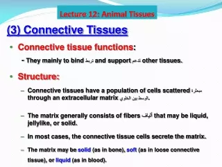
Connective Tissues The connective tissues are the gluing material that bound the different Structures together. A connective tissue consists of the different types of cells, fibers and ground substance. The fibers are embedded in the ground substance, now ground substance along fibers is called as Matrix
Connective Cells I – Fixed Connective tissues Cells Fibrocytes (or fibroblasts) Fat cells (Adipocytes) II - wandering cells. Histocytes (Macrophages) Monocytes Lymphocytes Plasma cells Eosinophils Mast cells
Types of Connective Tissue Fibers • Collagen Fibers: most widely distributed in the body formed by the Fibroblasts • Reticular Fibers mostly present in Basement membrane and capsules of different organs • Elastic Fibers are mainly found where the Elasticity is required
Collagen Fibers • Collagen fibers are also called as white fibers. They are arranged in the bundles with average length of 10 to 100 mm, the individual fiber has average diameter of 1 to 2 mm. when each collagen fiber examine under high magnification, it consists of thin collagen fibrils with of diameter 0.2 to 0.5 mm. The collagenous fibrils are arranged parallel, giving appearance of striation.
Structure of Collagen Fibers On Electron Microscopic examination revealed that each collagenous fibril consists still thinner microfilament with average length on 300 nm and diameter of 3 to 15 nm. Each microfilament is formed the macromolecule which is called as the Tropocollagen. Each tropocollagen consists of three polypeptide chains interwined together in form of a triple helix. The polypeptide chain is called is called an alpha chain.
Striations • The microfilaments arranged together to form collagenous fibrils. The tropocollagen arranged in such way that they are parallel to each other and they lying side by side, they are arranged in staggered fashion. One molecule is overlapped by the molecule by its one quarter length. The space between two adjacent is 40 nm
64 nm Banding This arrangement gives a typical appearance that after each 64 nm, there is light band, followed by dark band. This will give typical cross striations in some of collagen fibers. This is called as 64 nm periodicity or 64 nm banding under EM Microscope.
Types of Collagen Fibers There are many different tropocollagen types around (currently named type I to XXI). These types differ in their content of the amino acids hydroxyproline and hydroxylysine. They also differ in the amount of carbohydrates attached to the collagen molecules. The different types of tropocollagen give the fibers the structural and functional features which are appropriate for the organ in which the fibers are found.
Types of collagen • Collagen occurs in many places throughout the body. There are 28 types of collagen described in literature. Over 90% of the collagen in the body, however, are Collagens I, II, III, and IV. A simple way to remember their general functions is: • Collagen One - Bones (main component of bone) • Collagen Two - Cartilages (main component of cartilage) • Collagen Three - Reticular (main component of reticular fibers) • Collagen Four - forms the basement membrane
Reticular fibers or reticulin Reticular fibers or reticulin is a histological term used to describe a type of structural fiber composed of type III collagen. Reticular fibers crosslink to form a fine meshwork (reticulum). This network acts as a supporting mesh in soft tissues such as liver, bone marrow, and the tissues and organs of the lymphatic system
Structure Reticular fiber one or more types of very thin and delicately woven strands of type III collagen, these strands build a highly ordered cellular network and provide a supporting network. Many of these types of collagen have been combined with carbohydrate. Thus, they react with silver stains and with periodic acid-Schiff reagent but are not demonstrated with ordinary histological stains such as those using hematoxylin.
Formation & Distributions • Secreted by the fibroblast and Reticular Cells • Dermis of Skin, connective sheath surrounding the muscle, blood vessels, intestine, uterus, urinary bladder The reticular fibers are also found spleen, thymus, lymph node and Tonsils
Elastic fibers (or yellow fibers) are bundles of proteins (elastin) found in connective tissue and produced by fibroblasts and smooth muscle cells in arteries. These fibers can stretch up to 1.5 times their length
Formation of Elastic Fibers Elastic fibers, as the name suggests, are highly elastic and stretch in response to tension. In particular they are formed from the protein elastin. The amino acid composition of elastin, similar to collagen, is rich in glycine and proline, but in addition has two unusual amino acids, desmosine and isodesmosine. Elastic fibers also have a high content of valine.
Elastic Fibers • Elastic fibers, as the name suggests, are highly elastic and stretch in response to tension. In particular they are formed from the protein elastin. The amino acid composition of elastin, similar to collagen, is rich in glycine and proline, but in addition has two unusual amino acids, desmosine and isodesmosine. Elastic fibers also have a high content of valine.
Structure of Elastic Fibers The Elastic fibers are highly elastic and can easily be stretched by weak traction force and retrun to its original length when these forces are removed. Elastic fibers are seen in unstained preparation of loose connective tissues as very thin highly retractile strands with 0.2 to 1.0 mm, which branched and rejoined to form a loose network. In locations where elastic fibers are abundant they will produce yellow color to tissues.
Staining Technique In ordinary H & E stained tissue section the elastic fibers can not be identified easily because they are very faintly eosinophilic. But they are certain stain (for e.g. Resorcin-fuchsin technique for Elastic Tissue) can demonstrate the elastic fibers.
Development Electron Microscopic studies reveal that, during their formation the elastic fibers pass through three successive stages which can be both in embryonic and adult tissues. In initial stage the fibers consists of thin microfibrils, which are composed of a glycoprotein called fibrillin. In the next stage, an amorphous substance of low electron density appears in the center of developing fibers. This is called elastin. In the third stage of elastic fibers development, the amorphous elastin component constitutes more than 90% of fibers.
Distributions The elastic fibers are found in the ground substance of connective tissues throughout the body. For e.g. the lung, large size arteries, and certain ligaments
Ground substance • Ground substance is a term for the non-collagenous components of extracellular matrix. Cells are surrounded by extracellular matrix in tissues, which acts as a support for the cells. Ground substance traditionally does not include collagen but does include all the other proteinaceous components, including proteoglycans, matrix proteins and, most prevalent, water. • Glycosaminoglycans • sulfated variety includes chondroitin, dermatan, keratan, and heparan • non-sulfated • hyaluronic acid
Proteoglycan Molecules of these sulfated glycosaminoglycans become covalently attached as side chains to an axial core protein. The resulting structure, termed a proteoglycan, chondroitin-4-sulfate, chondroitin-6-sulfate, dermatan sulfate, heparan sulfate, heparin
Adhesive glycoproteins Help hold tissue structures together much like mortar binds bricks. One of these compounds, fibronectin, links the cytoskeleton of cells to collagen fibers and other elements of the extracellular matrix. Laminin helps hold epithelial cells to the basement membrane. Adhesive glycoproteins contribute to the ability of individual muscle fibers to work as a unit by holding them together when they contract. Other members of the adhesive glycoprotein family play an important role in the differentiation and growth of cells, development of blood vessels, and cell movement.
Basement membranes Basement membrane is a structure that supports an overlying epithelium or endothelium. Keratinocytes, glandular cells, and endothelial cells reside on basement membranes. Basement membrane consists of an electron dense membrane called basal lamina, about 300-700 angstrom in thickness, and an underlying network of reticular collagen fibrils which average 300 angstrom in diameter. This network is 0.1-2 micron in thickness.
The three parts of basement membranes The lamina lucida Above the lamina densa, next to the cell membrane, is the lamina lucida. This electron-lucent layer is traversed by delicate cords that extend from the lamina densa to the overlying cell membrane. These cords are thought to be composed, in part, of the extracellular portions of cell adhesion molecules (integrins).
The lamina densa • The lamina densa • The middle part, called the lamina densa, was formerly called the basal lamina. This layer consists of laminin-5, type IV collagen, which lacks the axial periodicity seen in other types of collagen, and proteoglycans. The lamina densa appears to be a product of the cells to which the basement membrane is attached (e.g., epithelial cells).
lamina fibroreticularis Below the lamina densa, next to the connective tissue, is the lamina fibroreticularis. This electron-lucent layer consists largely of reticular fibers (type III collagen) produced by fibroblasts in the underlying connective tissue. There is also some type VII collagen here.
Uploaded By.. M.FarrukhFayyaz