Download
1 / 27
270 likes | 423 Views
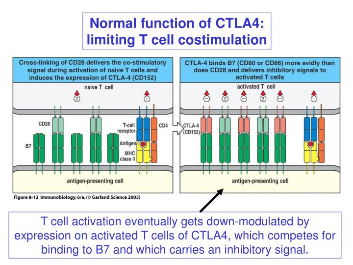
Normal function of CTLA4: limiting T cell costimulation. T cell activation eventually gets down-modulated by expression on activated T cells of CTLA4, which competes for binding to B7 and which carries an inhibitory signal. CTLA4-deficient mice.

E N D
Normal function of CTLA4: limiting T cell costimulation T cell activation eventually gets down-modulated by expression on activated T cells of CTLA4, which competes for binding to B7 and which carries an inhibitory signal.
CTLA4-deficient mice • Mice die at 3-4 weeks of systemic T cell activation and infiltration of multiple organs • Elevated antibodies, and autoantibodies
Absence of costimulation leads to T cell tolerance T cells encountering antigen on non-activated professional antigen presenting cells become inactivated inactivated
Self antigens perceived in the context of costimulation fail to prevent anergy and lead to stimulation • Antigens recognized with low affinity or those that are not present in the thymus do not lead to thymic deletion. • But T cells that recognize self antigen in the periphery can be tolerized in the absence of costimulation. • On the other hand, induction of costimulation by inflammation can break self-tolerance. • Classic example: original rabies vaccine. Vaccine was made from ground up spinal cords containing rabies virus. Immunized individuals developed neurological disease. • The same thing happens when normal spinal cord tissue is injected with adjuvant.
Disease can also be induced by immunizing (in adjuvant) with a single peptide from myelin basic protein Induction of EAE(experimental allergic encephalitis), a model for multiple sclerosis Immunization with many self antigens can lead to autoimmunity, depending on 1) the "strength" of adjuvant 2) genetics of the mouse (MHC plus other genes)
CD4 T cells drive the pathogenesis of EAE Isolate CD4 T cells
Regulatory T cells • Discovered relatively recently, though hints were in the scientific literature for decades. • Cells carry CD4 and CD25, along with TCRab. • Regulatory T cells are able to inhibit T cell responses by interacting with APCs at the same time as antigen reactive T cells. • While the mechanism of action is not well understood, there are several compelling reasons to believe that regulatory T cells may play an important role in preventing autoimmunity.
Regulatory T cells IL10 TGFb others?
Regulatory T cells, cont. • Mice thymectomized at day 3 of life have T cells, but they develop multiorgan autoimmune disease. • Rats or mice depleted of certain mature CD4 T cell subsets (CD4+CD25+ in mice) develop inflammatory bowel disease. • In both cases, reintroduction of the "missing" CD4 T cell subset (CD25+) leads to prevention of disease. • Humans and mice mutant in the transcription factor Fox3P develop a severe autoimmune disease. In humans IPEX (immunodysregulation, polyendocrinopathy, and enteropathy, X-linked). In mice, scurfy mutation. • Fox3P is currently the definitive marker for regulatory T cells and may be a master regulator of their development.
How are regulatory T cells (Tregs) made and educated? • Tregs require the thymus for development. • Tregs appear to be positively selected, but bind self peptide with intermediate affinity (i.e., too little to be negatively selected, but with higher affinity than the major T cell populations that are positively selected). • The current hope is that manipulating Tregs may allow treatment of existing autoimmune disease in an antigen specific way.
Epidemiology of autoimmunity Genetics - Many genetic loci affect the predisposition to autoimmune disease. By far the most important is the HLA locus Gender - In specific autoimmune diseases (SLE, MS), females are more commonly affected Environment - Studies in identical twins shows that there is >20% concordance in monozygotic twins, and < 5% in dizygotic twins
The strongest genetic association with disease maps to the MHC Type I diabetes (Protection)
Suceptibility to insulin dependent diabetes (IDDM) appears to be strongly increase in people carrying a particular HLA-DQ chain combination: the a chain of DQ8 and the b chain of DQ2. The nature of the key autoantigen is under intensive investigation.
Role of the environment Celiac disease is caused by T cell response to food peptide linked to a self protein Peptide linked to self protein, Transglutaminase Contains altered peptide
Celiac disease T cells also help autoreactive B cells that see transglutaminase-coupled to gluten peptide. Antibodies to transglutaminase are diagnostic of celiac disease.
Rheumatoid arthritis Autoantigen unknown
Figure 11-13 Anti-TNF therapy of rheumatoid arthritis
Some autoimmune diseases are caused by cross reactivity between microbial and self antigens
Clonal ignorance Express a flu viral protein as a transgene in the pancreas under the insulin promoter regulation. Mice are tolerant of the pancreatic beta cells until challenge with the virus, at which time they attack the pancreas.