Download
1 / 88
1.73k likes | 3.26k Views
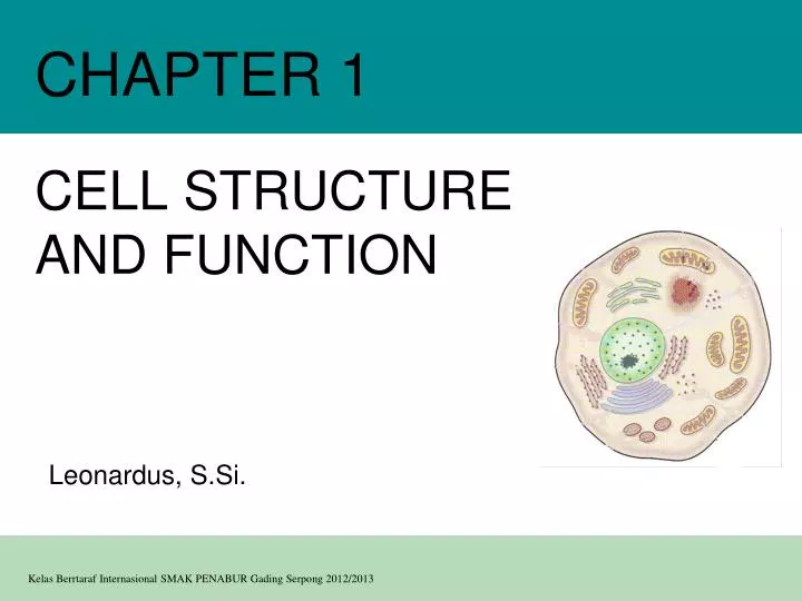
CHAPTER 1. CELL STRUCTURE AND FUNCTION. Leonardus , S.Si . Content . The microscope in cell studies Cells as the basic units of living organisms Detailed structure of typical animal and plant cells, as seen under the electron microscope

E N D
CHAPTER 1 CELL STRUCTURE AND FUNCTION Leonardus, S.Si.
Content • The microscope in cell studies • Cells as the basic units of living organisms • Detailed structure of typical animal and plant cells, as seen under the electron microscope • Outline functions of organelles in plant and animal cells • Characteristics of prokaryotic and eukaryotic cells
Learning Outcomes Candidates should be able to: • (a) [PA] use an eyepice graticule and stage micrometer to measure cells and be familiar with units (millimetre, micrometre, nanometre) used in cell studies; • (b) explain and distinguish between resolution and magnification, with reference to light microscopy and electron microscopy;
Learning Outcomes • (c) describe and interpret drawings and photographs of typical animal and plant cells, as seen under the electron microscope, recognising the following membrane systems and organelles: rough and smooth endoplasmic reticula, Golgi apparatus, mitochondria, ribosomes, lysosomes, chloroplasts, plasma/cell surface membrane, nuclear envelope, centrioles, nucleus and nucleolus; • (d) outline the functions of the membrane systems and organelles listed in (c);
Learning Outcomes • (e) [PA] compare and contrast the structure of typical animal and plant cells; • (f) [PA] draw and label low power diagrams of tissues (including a transverse section of a stems, roots, and leaves) and calculate the linear magnification of drawings; • (g) describe the structure of a prokaryotic cell and compare and contrast the structure of prokaryotic cells with eukaryotic cells; • (h) use the knowledge gained in this section in new situations or to solve related problems.
Introduction Overview: The Importance of Cells • All organisms are made of cells • The cell is the simplest collection of matter that can live • Cell structure is correlated to cellular function
Brief history • R. Hooke (1665), an English scientist: cell • A. v. Leeuwenhoek (1675), a Dutch lens maker: microscope, in the 150 years after his work, microscope lenses improved and scientists were able to observe and understand more parts of the cell.
Brief history • In the mid-19th century, three different scientists working separately each published important conclusions about cells. 1. In 1838, Dutch botanist Matthias Schleiden concluded that all plants are composed of cells. 2. One year later, a German zoologist by the name of Theodor Schwaan postulated that animals are also composed of cells. 3. And in 1855, Rudolph Virchow, a German doctor, asserted that all cells must come from other cells by the process of cell division.
Cell Theory (based on 200+ years of discoveries) • A. all living things are composed of cells • B. cells are the basic unit of structure & function of all living things • C. new cells are produced from existing cells
Why cells? • Because cells are the most basic unit of life that all organisms share, understanding cells is the key to learning about life itself.
Microscope • Cell biology: study of cells: cytology • To study cells, biologists use microscopes as the tools • Scientists use microscopes to visualize cells too small to see with the naked eye • Type of microscopes: • light microscope (uses light as a source of radiation) • electron microscope (uses electrons)
Microscope • Light microscopes (LM) • Pass visible light through a specimen • Magnify cellular structures with lenses
RESULT TECHNIQUE (a) Brightfield (unstained specimen). Passes light directly through specimen. Unless cell is naturally pigmented or artificially stained, image has little contrast. [Parts (a)–(d) show a human cheek epithelial cell.] 50 µm (b) Brightfield (stained specimen).Staining with various dyes enhances contrast, but most staining procedures require that cells be fixed (preserved). (c) Phase-contrast. Enhances contrast in unstained cells by amplifying variations in density within specimen; especially useful for examining living, unpigmented cells. Microscope • Use different methods for enhancing visualization of cellular structures
(d) (e) Fluorescence. Shows the locations of specific molecules in the cell by tagging the molecules with fluorescent dyes or antibodies. These fluorescent substances absorb ultraviolet radiation and emit visible light, as shown here in a cell from an artery. 50 µm (f) Confocal. Uses lasers and special optics for “optical sectioning” of fluorescently-stained specimens. Only a single plane of focus is illuminated; out-of-focus fluorescence above and below the plane is subtracted by a computer. A sharp image results, as seen in stained nervous tissue (top), where nerve cells are green, support cells are red, and regions of overlap are yellow. A standard fluorescence micrograph (bottom) of this relatively thick tissue is blurry. 50 µm Microscope Differential-interference-contrast (Nomarski). Like phase-contrast microscopy, it uses optical modifications to exaggerate differences in density, making the image appear almost 3D.
Microscope • Electron microscopes (EM) • Focus a beam of electrons through a specimen (TEM) or onto its surface (SEM)
TECHNIQUE RESULTS 1 µm Cilia (a) Scanning electron micro- scopy (SEM). Micrographs taken with a scanning electron microscope show a 3D image of the surface of a specimen. This SEM shows the surface of a cell from a rabbit trachea (windpipe) covered with motile organelles called cilia. Beating of the cilia helps move inhaled debris upward toward the throat. Microscope • The scanning electron microscope (SEM) • Provides for detailed study of the surface of a specimen
Longitudinal section of cilium Cross section of cilium (b) Transmission electron micro- scopy (TEM). A transmission electron microscope profiles a thin section of a specimen. Here we see a section through a tracheal cell, revealing its ultrastructure. In preparing the TEM, some cilia were cut along their lengths, creating longitudinal sections, while other cilia were cut straight across, creating cross sections. 1 µm Microscope • The transmission electron microscope (TEM) • Provides for detailed study of the internal ultrastructure of cells
Units of measurement in cell studies • International System of Units (SI Units)
10 m Human height 1 m Length of some nerve and muscle cells 0.1 m Light microscope Chicken egg 1 cm Frog egg 1 mm 100 µm Most plant and Animal cells Electron microscope 10 µ m NucleusMost bacteriaMitochondrion 1 µ m Electron microscope Smallest bacteria 100 nm Viruses 10 nm Ribosomes Proteins Lipids 1 nm Small molecules Atoms 0.1 nm Size of some biological structures Unaided eye Measurements 1 centimeter (cm) = 102 meter (m) = 0.4 inch 1 millimeter (mm) = 10–3 m 1 micrometer (µm) = 10–3 mm = 10–6 m 1 nanometer (nm) = 10–6 mm = 10–9 m
Measuring cells – eyepiece graticule eyepice graticule scale (arbitrary units) stage micrometer scale (marked in 0.01 mm and 0.1 mm divisions
Measuring cells – eyepiece graticule cheek cells on a slide on the stage of the microscope eyepiece graticule in the eyepiece of the microscopes
Magnification dan resolution • Magnification: the number of times larger an images is compared with the real size of the object size of image actual size of specimen • Resolution: the ability to distinguish between to separate points magnification =
Cell Types • Two types of cells make up every organisms • Prokaryotic • Eukaryotic
Cell Types • All cells have several basic features in common • They are bounded by a plasma membrane • They contain a semifluid substance called the cytosol • They contain chromosomes • They all have ribosomes
Prokaryotic cells • Prokaryotic cells • Do not contain a nucleus (do not have a nuclear membrane) • Have their DNA located in a region called the nucleoid
Pili: attachment structures on the surface of some prokaryotes Nucleoid: region where the cell’s DNA is located (not enclosed by a membrane) Ribosomes: organelles that synthesize proteins Plasma membrane: membrane enclosing the cytoplasm Cell wall: rigid structure outside the plasma membrane Capsule: jelly-like outer coating of many prokaryotes Bacterialchromosome 0.5 µm Flagella: locomotion organelles of some bacteria (b) A thin section through the bacterium Bacillus coagulans (TEM) (a) A typical rod-shaped bacterium Prokaryotic cells
Eukaryotic cells • Eukaryotic cells • Contain a true nucleus, bounded by a membranous nuclear envelope • Are generally quite a bit bigger than prokaryotic cells • Have extensive and elaborately arranged internal membranes, which form organelles • Have internal membranes that compartmentalize their functions
Eukaryotic cells: Plant and animal cell • A side-by-side view of an animal and plant cell. Plant cells contain special organelles called chloroplasts and an outer covering called a cell wall. Animal cells lack both of these structures.
Nuclear envelope ENDOPLASMIC RETICULUM (ER) NUCLEUS Nucleolus Rough ER Smooth ER Chromatin Flagellum Plasma membrane Centrosome CYTOSKELETON Microfilaments Intermediate filaments Ribosomes Microtubules Microvilli Golgi apparatus Peroxisome In animal cells but not plant cells: Lysosomes Centrioles Flagella (in some plant sperm) Lysosome Mitochondrion Animal cell
Nuclear envelope Rough endoplasmic reticulum Nucleolus NUCLEUS Chromatin Smooth endoplasmic reticulum Centrosome Ribosomes (small brwon dots) Central vacuole Tonoplast Golgi apparatus Microfilaments Intermediate filaments Microtubules Mitochondrion Peroxisome Plasma membrane Chloroplast Cell wall Plasmodesmata Wall of adjacent cell Plant cell CYTOSKELETON In plant cells but not animal cells: Chloroplasts Central vacuole and tonoplast Cell wall Plasmodesmata
Outside of cell Hydrophilic region TEM of a plasma membrane. The plasma membrane, here in a red blood cell, appears as a pair of dark bands separated by a light band. (a) Inside of cell 0.1 µm Hydrophobic region Hydrophilic region Phospholipid Proteins (b) Structure of the plasma membrane Plasma Membrane • Functions as a selective barrier • Allows food, oxygen, & water into the cell & waste products out of the cell. • Outer covering, protective layer around all cells Carbohydrate side chain back
The Nucleus: Genetic Library of the Cell • The nucleus • Contains most of the genes in the eukaryotic cell • Directs all cell activities • Contains instructions for everything the cell does • These instructions are found on a hereditary material called DNA • Usually the largest organelle back
Nucleus Nucleus 1 µm Nucleolus Chromatin Nuclear envelope: Inner membrane Outer membrane Nuclear pore Pore complex Rough ER Surface of nuclear envelope. 1 µm Ribosome 0.25 µm Close-up of nuclear envelope Nuclear lamina (TEM). Pore complexes (TEM). The Nuclear Envelope • Encloses the nucleus, separating its contents from the cytoplasm • controls movement of materials in & out of nucleus back
Nucleolus • “little nucleus” • Found in the nucleus back
Chromatin • contains genetic code that controls cell • made of DNA & proteins back
Ribosomes: Protein Factories in the Cell • Are particles made of ribosomal RNA and protein • Float freely or attached to the endoplasmic reticulum (ER) • made in the nucleolus • Carry out protein synthesis
Ribosomes Cytosol Free ribosomes Bound ribosomes Large subunit Small subunit 0.5 µm TEM showing ER and ribosomes Diagram of a ribosome Ribosomes: Protein Factories in the Cell ER Endoplasmic reticulum (ER) back
The Endoplasmic Reticulum: Biosynthetic Factory • The endoplasmic reticulum (ER) • Accounts for more than half the total membrane in many eukaryotic cells • A series of folded membranes that move materials (proteins) around in a cell • like a conveyor belt • Smooth ER – ribosomes not attached to ER • Rough ER – ribosomes attached to ER
The Endoplasmic Reticulum: Biosynthetic Factory • The function of smooth ER • Synthesizes lipids • Metabolizes carbohydrates • Stores calcium • Detoxifies poison
The Endoplasmic Reticulum: Biosynthetic Factory • The function of rough ER • Produces proteins and membranes, which are distributed by transport vesicles
Smooth ER Nuclear envelope Rough ER ER lumen Cisternae Ribosomes Transitional ER Transport vesicle 200 µm Smooth ER Rough ER The Endoplasmic Reticulum: Biosynthetic Factory back
The Golgi Apparatus: Shipping and Receiving Center • The Golgi apparatus • Receives many of the transport vesicles produced in the rough ER • Consists of flattened membranous sacs called cisternae • Functions of the Golgi apparatus include: • Sort and package proteins
cis face (“receiving” side of Golgi apparatus) 5 3 4 6 1 2 Vesicles coalesce to form new cis Golgi cisternae Vesicles move from ER to Golgi 0.1 0 µm Vesicles also transport certain proteins back to ER Cisternae Cisternal maturation: Golgi cisternae move in a cis- to-trans direction Vesicles form and leave Golgi, carrying specific proteins to other locations or to the plasma mem- brane for secretion trans face (“shipping” side of Golgi apparatus) Vesicles transport specific proteins backward to newer Golgi cisternae The Golgi Apparatus: Shipping and Receiving Center Golgi apparatus back TEM of Golgi apparatus
Lysosomes: Digestive Compartments • A lysosome • Is a membranous sac of hydrolytic enzymes • Can digest all kinds of macromolecules such as food or break down the cell when it dies • Lysosomes carry out intracellular digestion by • Phagocytosis • Autophagy
1 µm Nucleus Lysosome Lysosome contain active hydrolytic enzymes Food vacuole fuses with lysosome Hydrolytic enzymes digest food particles Digestive enzymes Lysosome Plasma membrane Digestion Food vacuole (a) Phagocytosis: lysosome digesting food Lysosomes: Phagocytosis