Download
1 / 69
690 likes | 706 Views
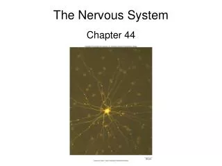
Chapter 12: Nervous System Cells. INTRODUCTION. The function of the nervous system, along with the endocrine system, is to communicate The nervous system consists of the brain, spinal cord, and nerves (Figure 12-1). ORGANIZATION OF THE NERVOUS SYSTEM.

E N D
INTRODUCTION The function of the nervous system, along with the endocrine system, is to communicate The nervous system consists of the brain, spinal cord, and nerves (Figure 12-1)
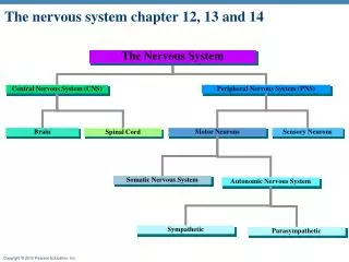
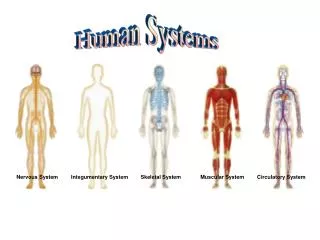
ORGANIZATION OF THE NERVOUS SYSTEM Subdivided into smaller “systems” by location (Figure 12-2) Central nervous system (CNS) Structural and functional center of the entire nervous system Consists of the brain and spinal cord Integrates sensory information, evaluates it, and initiates an outgoing response Peripheral nervous system (PNS) Nerves that lie in the “outer regions” of the nervous system Cranial nerves originate from the brain Spinal nerves originate from the spinal cord
ORGANIZATION OF THE NERVOUS SYSTEM (cont.) Afferent and efferent divisions Afferent division consists of all incoming sensory pathways Efferent division consists of all outgoing motor pathways “Systems” according to organs innervated Somatic nervous system Somatic motor division carries information to the somatic effectors (skeletal muscles) Somatic sensory division carries feedback information to somatic integration centers in the CNS
ORGANIZATION OF THE NERVOUS SYSTEM (cont.) • “Systems” according to organs innervated (cont.) • Autonomic nervous system (ANS) • Efferent division of ANS carries information to the autonomic or visceral effectors (smooth and cardiac muscles and glands) • Sympathetic division: prepares the body to deal with immediate threats to the internal environment; produces fight-or-flight response • Parasympathetic division: coordinates the body’s normal resting activities; sometimes called the rest-and-repair division
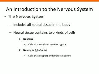
CELLS OF THE NERVOUS SYSTEM Glia (neuroglia) Glial cells support the neurons Five major types of glia (Figure 12-3) Astrocytes (in CNS) Star shaped; largest and most numerous type of glia Cell extensions connect to both neurons and capillaries Astrocytes transfer nutrients from the blood to the neurons Form tight sheaths around brain capillaries, which, with tight junctions between capillary endothelial cells, constitute the blood-brain barrier Microglia (in CNS) Small, usually stationary cells In inflamed brain tissue, they enlarge, move about, and carry on phagocytosis
CELLS OF THE NERVOUS SYSTEM (cont.) Five major types of glia (cont.) Ependymal cells (in CNS) Resemble epithelial cells and form thin sheets that line fluid-filled cavities in the CNS Some produce fluid; others aid in circulation of fluid Oligodendrocytes (in CNS) Smaller than astrocytes with fewer processes Hold nerve fibers together and produce the myelin sheath
CELLS OF THE NERVOUS SYSTEM (cont.)4th stopped here Five major types of glia (cont.) Schwann cells (in PNS) Found only in peripheral neurons Support nerve fibers and form myelin sheaths (Figure 12-4) Myelin sheath gaps are often called nodes of Ranvier Neurilemma is formed by cytoplasm of Schwann cell (neurolemmocyte) wrapped around the myelin sheath; essential for nerve regrowth Neuronal sheath is the myelin sheath plus the neurilemma (the whole Schwann wrapping around the axon) Satellite cells are Schwann cells that cover and support cell bodies in the PNS
CELLS OF THE NERVOUS SYSTEM (cont.) Neurons Excitable cells that initiate and conduct impulses that make possible all nervous system functions Components of neurons (Figure 12-5) Cell body (perikaryon) Ribosomes, rough endoplasmic reticulum, Golgi apparatus Provide protein molecules (neurotransmitters) needed for transmission of nerve signals from one neuron to another Neurotransmitters are packaged into vesicles Provide proteins for maintaining and regenerating nerve fibers Mitochondria provide energy (adenosine triphosphate) for neuron; some are transported to end of axon
CELLS OF THE NERVOUS SYSTEM (cont.) Components of neurons (cont.) Dendrites Each neuron has one or more dendrites, which branch from the cell body Conduct nerve signals to the cell body of the neuron Distal ends of dendrites of sensory neurons are receptors Dendritic spines: small, knoblike protrusions on dendrites of some brain neurons; serve as connection points for axons of other neurons Axon A single process extending from the axon hillock, sometimes covered by a fatty layer called a myelin sheath (Figure 12-6) Conducts nerve impulses away from the cell body of the neuron Distal tips of axons are telodendria, each of which terminates in a synaptic knob Axon varicosities: swellings that make contact (synapse) with other cells
CELLS OF THE NERVOUS SYSTEM (cont.) Components of neurons (cont.) Cytoskeleton Microtubules and microfilaments, as well as neurofibrils (bundles of neurofilaments) Allows the rapid transport of small organelles (Figure 12-7) Vesicles (some containing neurotransmitters), mitochondria Motor molecules shuttle organelles to and from the far ends of a neuron Functional regions of the neuron (Figure 12-8) Input zone: dendrites and cell body Summation zone: axon hillock Conduction zone: axon Output zone: telodendria and synaptic knobs of axon
CELLS OF THE NERVOUS SYSTEM (cont.) Classification of neurons Structural classification: according to number of processes extending from cell body (Figure 12-9) Multipolar: one axon and several dendrites Bipolar: only one axon and one dendrite; least numerous kind of neuron Unipolar (pseudounipolar): one process comes off neuron cell body but divides almost immediately into two fibers: central fiber and peripheral fiber
CELLS OF THE NERVOUS SYSTEM (cont.) • Classification of neurons (cont.) • Functional classification (Figure 12-10) • Afferent (sensory) neurons: conduct impulses to spinal cord or brain • Efferent (motor) neurons: conduct impulses away from spinal cord or brain toward muscles or glandular tissue
CELLS OF THE NERVOUS SYSTEM (cont.) Reflex arc A signal conduction route to and from the CNS, with the electrical signal beginning in receptors and ending in effectors Three-neuron arc most common; consists of afferent neurons, interneurons, and efferent neurons (Figure 12-11) Afferent neurons conduct impulses to the CNS from the receptors Efferent neurons conduct impulses from the CNS to effectors (muscle or glandular tissue) Two-neuron arc is simplest form; consists of afferent and efferent neurons Synapse Where nerve signals are transmitted from one neuron to another Two types: electrical and chemical; chemical synapses are typical in the adult
NERVES AND TRACTS Nerves: bundles of peripheral nerve fibers held together by several layers of connective tissue (Figure 12-12) Endoneurium: delicate layer of fibrous connective tissue surrounding each nerve fiber Perineurium: connective tissue holding together fascicles (bundles of fibers) Epineurium: fibrous coat surrounding numerous fascicles and blood vessels to form a complete nerve
NERVES AND TRACTS (cont.) White matter PNS: myelinated nerves CNS: myelinated tracts Gray matter Composed of cell bodies and unmyelinated fibers CNS: referred to as nuclei PNS: referred to as ganglia Mixed nerves Contain sensory and motor neurons Sensory nerves have predominantly sensory neurons Motor nerves have predominantly motor neurons
REPAIR OF NERVE FIBERS Mature neurons are incapable of cell division; therefore damage to nervous tissue can be permanent Neurons have limited capacity to repair themselves If the damage is not extensive, the cell body and neurilemma are intact, and scarring has not occurred, nerve fibers can be repaired
REPAIR OF NERVE FIBERS (cont.) Stages of repair of an axon in a peripheral motor neuron (Figure 12-13) After injury, distal portion of axon and myelin sheath degenerates Macrophages remove the debris Remaining neurilemma and endoneurium form a tunnel from the point of injury to the effector New Schwann cells grow in tunnel to maintain a path for axon regrowth Cell body reorganizes its Nissl bodies to provide the needed proteins to extend the remaining healthy portion of the axon Axon “sprouts” appear When sprout reaches tunnel, its growth rate increases Skeletal muscle cell atrophies until nervous connection is reestablished In CNS, similar repair of damaged nerve fibers is unlikely
NERVE IMPULSES Membrane potentials All living cells maintain a difference in the concentration of ions across their membranes Membrane potential: slight excess of positively charged ions on the outside of the membrane and slight deficiency of positively charged ions on the inside of the membrane (Figure 12-14)
NEUROTRANSMITTERS Neurotransmitters: means by which neurons communicate with one another; more than 30 compounds are known to be neurotransmitters, and dozens of others are suspected
NEUROTRANSMITTERS (cont.) Amino acids Believed to be among the most common neurotransmitters of the CNS In the PNS, amino acids are stored in synaptic vesicles and used as neurotransmitters Other small-molecule transmitters Nitric oxide derived from an amino acid Nitric oxide from a postsynaptic cell signals the presynaptic neuron, providing feedback in a neural pathway
NERVE IMPULSES contd • Difference in electrical charge is called potential because it is a type of stored energy • Polarized membrane: a membrane that exhibits a membrane potential • Magnitude of potential difference between the two sides of a polarized membrane is measured in volts (V) or millivolts (mV); the sign of a membrane’s voltage indicates the charge on the inside surface of a polarized membrane
NERVE IMPULSES (cont.) Resting membrane potential Membrane potential maintained by a nonconducting neuron’s plasma membrane; typically 70 mV The slight excess of positive ions on a membrane’s outer surface is produced by ion transport mechanisms and the membrane’s permeability characteristics The membrane’s selective permeability characteristics help maintain a slight excess of positive ions on the outer surface of the membrane (Figure 12-15) Sodium-potassium pump (Figure 12-16) Active transport mechanism in plasma membrane that transports sodium (Na+) and potassium (K+) ions in opposite directions and at different rates Maintains an imbalance in the distribution of positive ions, resulting in the inside surface becoming slightly negative compared with its outer surface
NERVE IMPULSES (cont.) Local potentials Local potentials: slight shift away from the resting membrane in a specific region of the plasma membrane (Figure 12-17) Excitation: when a stimulus triggers the opening of additional Na+ channels, allowing the membrane potential to move toward zero (depolarization) Inhibition: when a stimulus triggers the opening of additional K+ channels, increasing the membrane potential (hyperpolarization) Local potentials are called graded potentials because the magnitude of deviation from the resting membrane potential is proportional to the magnitude of the stimulus
ACTION POTENTIAL Action potential: the membrane potential of a neuron conducting an impulse; also known as a nerve impulse Mechanism that produces the action potential (Figures 12-18 and 12-19) When an adequate stimulus triggers stimulus-gated Na+ channels to open, allowing Na+ to diffuse rapidly into the cell, which produces a local depolarization As threshold potential is reached, voltage-gated Na+ channels open and more Na+ enters the cell, causing further depolarization The action potential is an all-or-none response Voltage-gated Na+ channels stay open for only about 1 ms before they automatically close
ACTION POTENTIAL (cont.) After action potential peaks, membrane begins to move back toward the resting membrane potential when K+ channels open, allowing outward diffusion of K+; process is known as repolarization Brief period of hyperpolarization occurs, then the resting membrane potential is restored by the Na-K pumps Refractory period (Figure 12-20) Absolute refractory period: brief period (lasting approximately 0.5 ms) during which a local area of a neuron’s membrane resists restimulation and will not respond to a stimulus, no matter how strong Relative refractory period: time when the membrane is repolarized and restoring the resting membrane potential; the few milliseconds after the absolute refractory period; will respond only to a very strong stimulus
Membrane potential: slight excess of positively charged ions on the outside of the membrane and slight deficiency of positively charged ions on the inside of the membrane • Sodium-potassium pump Maintains an imbalance in the distribution of positive ions, resulting in the inside surface becoming slightly negative compared with its outer surface • Action potential: the membrane potential of a neuron conducting an impulse; also known as a nerve impulse • Local potentials: slight shift away from the resting membrane in a specific region of the plasma membrane (Figure 12-17) • Excitation: when a stimulus triggers the opening of additional Na+ channels, allowing the membrane potential to move toward zero (depolarization) • Inhibition: when a stimulus triggers the opening of additional K+ channels, increasing the membrane potential (hyperpolarization)
After action potential peaks, membrane begins to move back toward the resting membrane potential when K+ channels open, allowing outward diffusion of K+; process is known as repolarization • Brief period of hyperpolarization occurs, then the resting membrane potential is restored by the Na-K pumps • Refractory period (Figure 12-20) • Absolute refractory period: brief period (lasting approximately 0.5 ms) during which a local area of a neuron’s membrane resists restimulation and will not respond to a stimulus, no matter how strong
ACTION POTENTIAL (cont.) Conduction of the action potential At the peak of the action potential, the plasma membrane’s polarity is now the reverse of the resting membrane potential The reversal in polarity causes electrical current to flow between the site of the action potential and the adjacent regions of membrane and triggers voltage-gated Na+ channels in the next segment to open; this next segment exhibits an action potential (Figure 12-21) This cycle continues to repeat The action potential never moves backward because of the refractory period In myelinated fibers, action potentials in the membrane only occur at the nodes of Ranvier; this type of impulse conduction is called saltatory conduction (Figure 12-22) Speed of nerve conduction depends on diameter and on the presence or absence of a myelin sheath