Download
1 / 16
170 likes | 709 Views
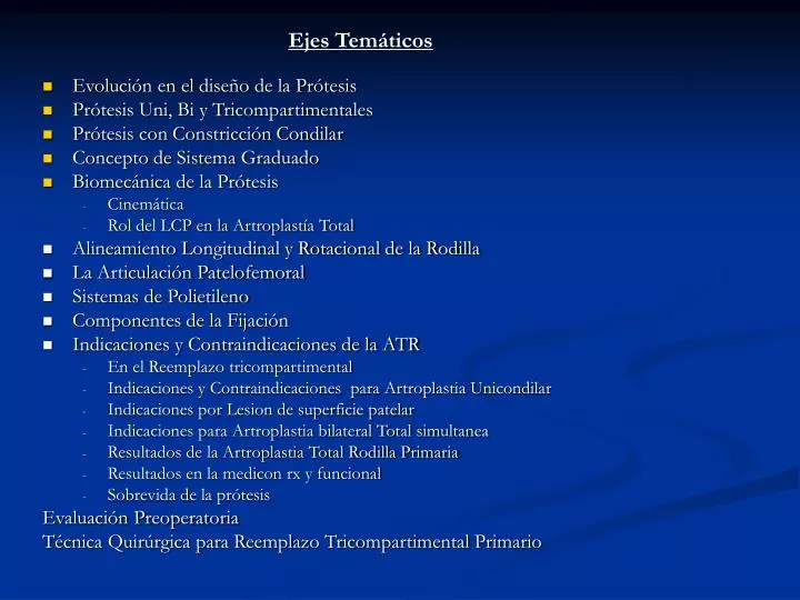
Ejes Temáticos. Evolución en el diseño de la Prótesis Prótesis Uni, Bi y Tricompartimentales Prótesis con Constricción Condilar Concepto de Sistema Graduado Biomecánica de la Prótesis Cinemática Rol del LCP en la Artroplastía Total Alineamiento Longitudinal y Rotacional de la Rodilla

E N D
Ejes Temáticos • Evolución en el diseño de la Prótesis • Prótesis Uni, Bi y Tricompartimentales • Prótesis con Constricción Condilar • Concepto de Sistema Graduado • Biomecánica de la Prótesis • Cinemática • Rol del LCP en la Artroplastía Total • Alineamiento Longitudinal y Rotacional de la Rodilla • La Articulación Patelofemoral • Sistemas de Polietileno • Componentes de la Fijación • Indicaciones y Contraindicaciones de la ATR • En el Reemplazo tricompartimental • Indicaciones y Contraindicaciones para Artroplastia Unicondilar • Indicaciones por Lesion de superficie patelar • Indicaciones para Artroplastia bilateral Total simultanea • Resultados de la Artroplastia Total Rodilla Primaria • Resultados en la medicon rx y funcional • Sobrevida de la prótesis Evaluación Preoperatoria Técnica Quirúrgica para Reemplazo Tricompartimental Primario
Abordaje Quirúrgico en Artroplastía Total Rodilla C. Fernando Villanueva Y. MR2 COT- UNMSM - HNDM
Incisión Fig. 6-34 Medial parapatellar retinacular approach
Disección de Planos Fig. 6-35 Medial capsule and deep portion of MCL are elevated subperiosteally. (Redrawn from Krackow KA: The technique of total knee arthroplasty, St Louis, 1990, Mosby.)
Disección de Planos Profundos Fig. 6-36 Lateral patellofemoral plicae are cut to allow mobilization of extensor mechanism. (Redrawn from Krackow KA: The technique of total knee arthroplasty, St Louis, 1990, Mosby.)
Disección de Planos Profundos Fig. 6-37 Subvastus approach involves lifting entire extensor mechanism off medial intermuscular septum and subluxing it laterally for exposure. (Redrawn from Insall JN: Surgical approaches. In Insall JN, ed: Surgery of the knee, ed 2, New York, 1993, Churchill Livingstone.)
Ejes de Alineamiento a nivel de linea intercondilea Fig. 6-39 Alignment axes in knee with normal condylar shape. Resection perpendicular to anteroposterior axis (AP) or parallel to epicondylar axis (epi) results in resection line (x) that is slightly externally rotated relative to posterior condylar axis (PC). This results in correct positioning of the femoral component. (From Arima J, Whiteside LA, McCarthy DS, White SE: J Bone Joint Surg 77A:1331, 1995.)
Disección de Planos Profundos Fig. 6-38 Midvastus approach shown as dotted line with right knee in 90 degrees flexion. (Redrawn from Engh GA, Parks NL: Clin Orthop 351:270, 1998.)
Sección de Componente Femoral Fig. 6-41 A, Chamfer cuts complete distal femoral resection in cruciate-retaining arthroplasty. B, Intercondylar notch cut to accommodate post and cam mechanism in cruciate-substituting arthroplasty. (Redrawn from Scott RD, Thornhill TS, Ranawat CS, et al: Primary cruciate-retaining procedure and primary cruciate-substituting procedure surgical technique manual, using the PFC modular total knee system, Raynam, Mass, 1994, Johnson & Johnson
Cortes de Componente Femoral conservando LCP Fig. 6-43 PCL may act as central tether and require lengthening or release. (Redrawn from Krackow KA: The technique of total knee arthroplasty, St Louis, 1990, Mosby.)
Reparación de LCM Fig. 6-44 Advancement of femoral origin of MCL and fixation in medial epicondyle with screw and washer. (Redrawn from Krackow KA: The technique of total knee arthroplasty, St Louis, 1990, Mosby.)
Sección de LCP Fig. 6-45 PCL can be "recessed" by releasing it from superior surface of tibia and proximal portion of its posterior tibial attachment. (Redrawn after Elizabeth Roselius in Insall JN: Surgical techniques and instrumentation in total knee arthroplasty. In Insall JN, ed: Surgery of the knee, ed 2, New York, 1993, Churchill Livingstone.)