Download
1 / 24
330 likes | 526 Views
EYE ANATOMY. EQ; What are the components of your eye?. Root Words. Eye Anatomy. Eye Anatomy. The orbital bone The eye socket Eye is cushioned within orbit by pads of fat Lacrimal gland Produces tears.

E N D
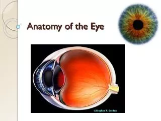
EYE ANATOMY EQ; What are the components of your eye?
Eye Anatomy http://everlastingelephants.blogspot.com/2009/08/what-is-eye-cataract.html
Eye Anatomy • The orbital bone • The eye socket • Eye is cushioned within orbit by pads of fat • Lacrimal gland • Produces tears http://commons.wikimedia.org/wiki/File:Eye_orbit_anatomy_anterior2.jpg http://mwsu-bio101.ning.com/forum/topics/distinct-human-celltypes-1?commentId=2263214%3AComment%3A10331
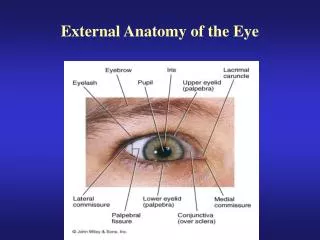
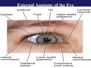
Eye Anatomy • Eyelids (L): • Protection against particles and light • Help spread tears over surface of eye- moist & comfort • Eyelashes (L): • Filter out foreign matter http://www.medical-look.com/human_anatomy/organs/Eyelids_and_eyelashes.html
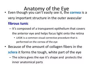
Eye Anatomy • Sclera (S): • “White of the eye” • Tough, opaque tissue that extends around the eye • Attached to the extraocular muscles http://www.thirdeyehealth.com/sclera.html
Eye Anatomy • Extraocular Muscles • Help move the eye left, right, up, down and diagonally • These 6 muscles are: • Superior rectus • Inferior rectus • Medial rectus • Lateral rectus • Inferior oblique • Superior oblique http://media.photobucket.com/image/introduction%20to%20eye%20anatomy/trimurtulu/Eye.jpg
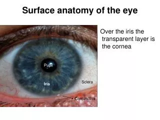
Eye Anatomy • Cornea (K): • Clear tissue in front of the Iris • Function: • Focus light as it enters eye • Avascular • Only organ that has no blood vessels http://commons.wikimedia.org/wiki/File:Cornea.jpg
Eye Anatomy • Pupil (P): • Central opening of iris • Iris (I): • Ring shaped tissue • Colored part of eye • Controls the amount of light that enters the eye • Two muscle fibers: • Contraction • Constricts pupil in bright light • Dilation • Dilates pupil in dark http://www.bioconsulting.com/Bio_Tech_Assessment.html http://www.goodhope.org.uk/departments/eyedept/angleclosureetc.htm
Eye Anatomy • Crystalline Lens: • Clear, flexible structure • Behind the iris & pupil • The lens & ciliary body help control fine focusing of light as it passes through the eye http://www.smartplanet.com/business/blog/smart-takes/artificial-lens-implant-to-give-patients-high-definition-vision-better-than-2020/2558/
Eye Anatomy • Vitreous Chamber: • Located behind the lens & in front of the retina • Filled with a gel-like fluid called the vitreous humor • The vitreous help maintain the shape of the eye http://www.ophthobook.com/questions/question-how-many-chambers-are-there-in-the-eye
Eye Anatomy • Retina: • Acts like the film in a camera to create an image • Converts light signals into nerve signal then send these signals to the optic nerve • Optic nerve carries the signals to the brain • Rods- low light situations • Cones- allows you to see color hhttp://www1.appstate.edu/~kms/classes/psy3203/EyePhysio/human_retina.htm http://www.answersingenesis.org/tj/v13/i1/retina.asp
Eye Anatomy • Optic Nerve • A bundle of 1 million nerve fibers • Responsible for transmitting nerve signals from the eye to the brain • The optic disc is the front surface of the optic nerve • The optic disc is visible on the retina http://cssd.us/body.cfm?id=802 http://www.wollongong.youronlinecommunity.com.au/wollongong-online/2008/50/walkthrulife/eye-health.html
Eye Anatomy • Macula • Located in the central part of the retina • Responsible for giving sharp central vision • Used for reading, recognizing faces, and watching TV http://www.dukehealth.org/eye_center/specialties/macular_degeneration/care_guides/macular_degeneration_frequently_asked_questions
Optic Chiasm The X-Shaped space infront of the pituitary gland where the optic nerves cross the brain
Ciliary Body Ciliary muscle, which changes the shape of the lens when your eyes focus on something. This process is called Accommodation.
tapetumlucidum - A layer of tissue lying behind the retina that reflex more light for the photoreceptors. - Found in nocturnal mammals for night vision
Cataract • Clouding of the lens which leads to decreased vision • Mostly due to age: degrading of the lens
Glaucoma • Disease that damages the optic nerve which results in blindness • Results from increased pressure from fluid build up
LEFT SIDE ACTIVITY • Draw and label the components of the eye. Describe the function of each of the components.