Download
1 / 43
430 likes | 433 Views
Learn about the location and structure of the kidneys, the process of urine formation, and the role of the urinary system in maintaining water balance and eliminating waste products.

E N D
Organs of the Urinary system • Kidneys • Ureters • Urinary bladder • Urethra Figure 15.1a
Elimination of waste products Nitrogenous wastes Toxins Drugs Regulate aspects of homeostasis Water balance Electrolytes Acid-base balance Blood pressure RBC production Activation of vit.D Functions of the Urinary System
Location of the Kidneys • Against the dorsal body wall • At the level of T12 to L3 • The right kidney is slightly lower than the left • Attached to ureters, renal blood vessels, and nerves at renal hilus • Atop each kidney is an adrenal gland
Coverings of the Kidneys • Renal capsule • Surrounds each kidney • Adipose capsule • Surrounds the kidney • Provides protection to the kidney • Helps keep the kidney in its correct location
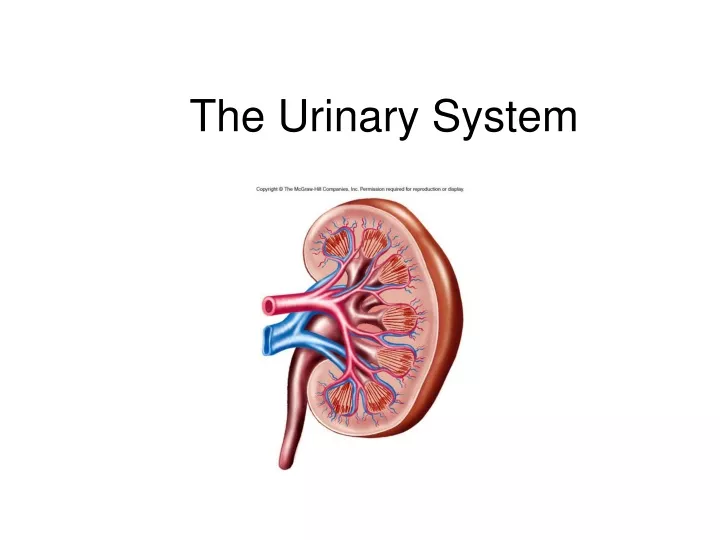
Regions of the Kidney • Renal cortex – outer region • Renal medulla – inside the cortex • Renal pelvis – inner collecting tube Figure 15.2b
Kidney Structures • Medullary pyramids – triangular regions of tissue in the medulla • Renal columns – extensions of cortex-like material inward • Calyces – cup-shaped structures that funnel urine towards the renal pelvis
Blood Flow in the Kidneys Figure 15.2c
Nephrons • The structural & functional units of the kidneys • Responsible for forming urine • Main structures of the nephrons • Glomerulus • Renal tubule
Glomerulus • A specialized capillary bed • Attached to arterioles on both sides (maintains high pressure) • Large afferent arteriole • Narrow efferent arteriole Figure 15.3c
Glomerulus • Capillaries are covered with podocytes from the renal tubule • The glomerulus sits within a glomerular capsule (the first part of the renal tubule) Figure 15.3c
Renal Tubule • Glomerular (Bowman’s) capsule • Proximal convoluted tubule • Loop of Henle • Distal convoluted tubule
Cortical nephrons Located entirely in the cortex Includes most nephrons Juxtamedullary nephrons Found at the boundary of the cortex and medulla Types of Nephrons Figure 15.3a
Urine Formation Processes • Filtration • Reabsorption • Secretion Figure 15.4
Filtration • Nonselective passive process • Water and solutes smaller than proteins are forced through capillary walls • Blood cells cannot pass out to the capillaries • Filtrate is collected in the glomerular capsule and leaves via the renal tubule
Reabsorption • The peritubular capillaries reabsorb several materials • Some water • Glucose • Amino acids • Ions • Some reabsorption is passive, most is active • Most reabsorption occurs in the proximal convoluted tubule
Materials Not Reabsorbed • Nitrogenous waste products • Urea • Uric acid • Creatinine • Excess water
Secretion – Reabsorption in Reverse • Some materials move from the peritubular capillaries into the renal tubules • Hydrogen and potassium ions • Creatinine • Materials left in the renal tubule move toward the ureter
Formation of Urine Figure 15.5
Characteristics of Urine Used for Medical Diagnosis • Colored somewhat yellow due to the pigment urochrome (from the destruction of hemoglobin) and solutes • Sterile • Slightly aromatic • Normal pH of around 6 • Specific gravity of 1.001 to 1.035
Ureters • Slender tubes attaching the kidney to the bladder • Continuous with the renal pelvis • Enter the posterior aspect of the bladder • Runs behind the peritoneum • Peristalsis aids gravity in urine transport
Urinary Bladder • Smooth, collapsible, muscular sac • Temporarily stores urine Figure 15.6
Urinary Bladder • Trigone – three openings • Two from the ureters • One to the urethrea Figure 15.6
Urinary Bladder Wall • Three layers of smooth muscle (detrusor muscle) • Mucosa made of transitional epithelium • Walls are thick and folded in an empty bladder • Bladder can expand significantly without increasing internal pressure
Urethra • Thin-walled tube that carries urine from the bladder to the outside of the body by peristalsis • Release of urine is controlled by two sphincters • Internal urethral sphincter (involuntary) • External urethral sphincter (voluntary)
Urethra Gender Differences • Length • Females – 3–4 cm (1 inch) • Males – 20 cm (8 inches) • Location • Females – along wall of the vagina • Males – through the prostate and penis
Urethra Gender Differences • Function • Females – only carries urine • Males – carries urine and is a passageway for sperm cells
Micturition (Voiding) • Both sphincter muscles must open to allow voiding • The internal urethral sphincter is relaxed after stretching of the bladder • Activation is from an impulse sent to the spinal cord and then back via the pelvic splanchnic nerves • The external urethral sphincter must be voluntarily relaxed
Maintaining Water Balance • Normal amount of water in the human body • Young adult females – 50% • Young adult males – 60% • Babies – 75% • Old age – 45% • Water is necessary for many body functions and levels must be maintained
Distribution of Body Fluid • Intracellular fluid (inside cells) • Extracellular fluid (outside cells) • Interstitial fluid • Blood plasma Figure 15.8
The Link Between Water and Salt • Changes in electrolyte balance causes water to move from one compartment to another • Alters blood volume and blood pressure • Can impair the activity of cells
Maintaining Water Balance • Water intake must equal water output • Sources for water intake • Ingested foods and fluids • Water produced from metabolic processes • Sources for water output • Vaporization out of the lungs • Lost in perspiration • Leaves the body in the feces • Urine production
Maintaining Water Balance • Dilute urine is produced if water intake is excessive • Less urine (concentrated) is produced if large amounts of water are lost • Proper concentrations of various electrolytes must be present
Regulation of Water and Electrolyte Reabsorption • Regulation is primarily by hormones • Antidiuretic hormone (ADH) prevents excessive water loss in urine • Aldosterone regulates sodium ion content of extracellular fluid • Triggered by the rennin-angiotensin mechanism • Cells in the kidneys and hypothalamus are active monitors
Maintaining Acid-Base Balance in Blood • Blood pH must remain between 7.35 and 7.45 to maintain homeostasis • Alkalosis – pH above 7.45 • Acidosis – pH below 7.35 • Most ions originate as byproducts of cellular metabolism
Maintaining Acid-Base Balance in Blood • Most acid-base balance is maintained by the kidneys • Other acid-base controlling systems • Blood buffers • Respiration
Blood Buffers • Molecules react to prevent dramatic changes in hydrogen ion (H+) concentrations • Three major chemical buffer systems • Bicarbonate buffer system • Phosphate buffer system • Protein buffer system
The Bicarbonate Buffer System • Mixture of carbonic acid (H2CO3) and sodium bicarbonate (NaHCO3) • Bicarbonate ions (HCO3–) react with strong acids to change them to weak acids • Carbonic acid dissociates in the presence of a strong base to form a weak base and water
Respiratory System Controls of Acid-Base Balance • Carbon dioxide in the blood is converted to bicarbonate ion and transported in the plasma • Increases in hydrogen ion concentration produces more carbonic acid • Excess hydrogen ion can be blown off with the release of carbon dioxide from the lungs • Respiratory rate can rise and fall depending on changing blood pH
Renal Mechanisms of Acid-Base Balance • Excrete bicarbonate ions if needed • Conserve or generate new bicarbonate ions if needed • Urine pH varies from 4.5 to 8.0
Developmental Aspects of the Urinary System • Functional kidneys are developed by the third month • Urinary system of a newborn • Bladder is small; urine cannot be concentrated • Control of the voluntary urethral sphincter starts until age 18 months • Urinary infections are the only common problems before old age
Aging and the Urinary System • There is a progressive decline in urinary function • The bladder shrinks with aging • Urinary retention is common in males