Download
1 / 26
270 likes | 335 Views
HYPOXIA AND CYANOSIS. Prof. Sultan Ayoub Meo MBBS, M.Phil, Ph.D (Pak), M Med Ed (Dundee), FRCP (London) FRCP (Dublin), FRCP (Glasgow), FRCP (Edinburgh) Professor and Consultant, Department of Physiology, College of Medicine, King Saud University, Riyadh, Saudi Arabia. HYPOXIA.

E N D
HYPOXIA AND CYANOSIS Prof. Sultan Ayoub Meo MBBS, M.Phil, Ph.D (Pak), M Med Ed (Dundee), FRCP (London) FRCP (Dublin), FRCP (Glasgow), FRCP (Edinburgh) Professor and Consultant, Department of Physiology, College of Medicine, King Saud University, Riyadh, Saudi Arabia
HYPOXIA Hypoxia is defined as an inadequate supply of oxygen to the body tissues. It can be classified in the following types. Hypoxic hypoxia Anemic hypoxia Stagnant hypoxia (Hypokinetic or Ischemic hypoxia) Histotoxic hypoxia
HYPOXIC HYPOXIA Hypoxic hypoxia is also known as arterial hypoxia. This is seen when there is a lack of oxygenation of blood in the lungs, which leads to a low PO2 in arterial blood. Since less amounts of Hb is converted to oxy-Hb. The tissues are supplied with blood deficient in oxygen. Hypoxic hypoxia can occur in the following conditions: • High altitude • Fluid in the lungs (pulmonary oedema) • Obstruction in the respiratory passages • Emphysema
HYPOXIC HYPOXIA a) Alveolar hypoventilation, e.g. • Reduced PO2 in inspired air (high altitude) • Increased airway resistance • Reduced lung compliance • Paralysis of respiratory muscles • Depressed respiratory centre b) Diffusion abnormalities: Impaired diffusion from alveolar to pulmonary capillary blood can lead to arterial hypoxia. It is seen in conditions like alveolar-capillary block.
HYPOXIC HYPOXIA c) ventilation-perfusion imbalance (including increased physiological dead space and physiological shunt): If ventilation and blood flow are mismatched in various parts of the lung, impairment of both oxygen and carbon dioxide diffusion results. Ventilation perfusion imbalance may be caused by uneven ventilation, e.g. obstructive lung conditions, or uneven perfusion, e.g. consolidation of the lung.
HYPOXIC HYPOXIA d) Right to left shunt: Blood passes from the systemic venous without going through the gas exchanging part of the lungs. This type of hypoxia can be differentiated clinically from other types by giving the subject 100% oxygen to breathe. Hypoxia because of the shunt will not be abolished while in other types PO2 in the arterial system will improve considerably.
ANEMIC HYPOXIA This condition is characterized by decreased oxygen carrying capacity of the blood due to decreased hemoglobin level. Anemic hypoxia is seen in haemorrhagic anemia [Decreased RBC / quality, or the failure of hemoglobin to carry its normal concentration of oxygen, as in carbon monoxide (CO) poisoning. Altered haemoglobin formation egmethaemoglobin, sulphaemoglobin, and carboxyhaemoglobin.
STAGNANT HYPOXIA Stagnant hypoxia [(Hypokinetic or Ischemic hypoxia] occurs in conditions in which there is a decreased rate of blood flow throughout the body or a part of the body. It may be caused by congestiveheart failure, circulatory shock and arteriosclerosis. General slowing of circulation [heart failure and shock] Local slowing: vasoconstriction, cold, arterial wall spasm.
HISTOTOXIC HYPOXIA In histotoxic hypoxia the tissue are unable to use oxygen even though plenty of oxygen is available. This is caused by inhibition of the tissue respiration electron transport chain. The best example is cyanide poisoning, where tissue cytochromeoxidases are knocked out and tissue is unable to utilize oxygen.
HYPOXIA AND CLINICAL FEATURES Signs and symptoms of hypoxia: Clinical features depend on how fast and how severely partial pressure of O2 is decreased. Fulminant hypoxia: This occurs very rapidly, within seconds. Unconsciousness occurs in 15-20 seconds and brain tissue death occurs in 4-5 minutes. Acute hypoxia: In acute hypoxia body reflexes are slowed, there may be slurred speech, unconsciousness, coma and death may occur. Chronic hypoxia: The symptoms of chronic hypoxia, fatigue, difficulty in breathing, and shortness of breath can occur. Signs of hypoxia: Cyanosis, Tachy-cardia and Tachypne
EFFECTS OF HYPOXIA ON THE BODY Effects of Hypoxia on the Body. Hypoxia, if severe enough, can cause death of cells throughout the body, but in less severe degrees it causes principally depressed mental activity, sometimes culminating in coma, Reduced work capacity of the muscles.
EFFECTS OF HYPOXIA *EXTRA SLIDE ON THE BODY At 12,000 feet, drowsiness, lassitude, mental and muscle fatigue, sometimes headache, occasionally nausea, and euphoria. These effects progress to a stage of twitchings or seizures above 18,000 feet and above 23,000 feet in the unacclimatized person, coma followed shortly thereafter by death. One of the most important effects of hypoxia is decreased mental proficiency, which decreases judgment, memory, and performance of discrete motor movements. if an unacclimatized person stays at 15,000 feet for 1 hour, mental proficiency ordinarily falls to about 50 per cent of normal, and after 18 hours at this level it falls to about 20 per cent of normal.
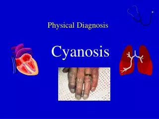
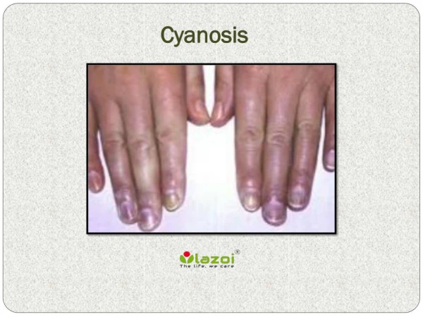
CYANOSIS The term cyanosis means blueness of the skin due to excessive amount of deoxygenated hemoglobin in the blood [skin blood vessels especially in the capillaries]. This deoxygenated hemoglobin has an intense dark blue-purple color that is transmitted through the skin. Cyanosis appears whenever the arterial blood contains more than 5 grams of deoxygenated hemoglobin in each 100 milliliters of blood.
CYANOSIS A person with anemia never becomes cyanotic because there is not enough hemoglobin for 5 grams to be deoxygenated in 100 milliliters of arterial blood. In a person with excess red blood cells as occurs in polycythemia the great excess of available hemoglobin that can become deoxygenated leads frequently to cyanosis.
CYANOSIS Causes of Cyanosis: i. Inadequate oxygenation of blood in the lungs • High altitude • Obstruction of respiratory passages • Pneumonconiosis • Emphysema • CO-poisoning ii. Presence of an aerated shunt between vessels • Coaractation of aorta • Fallotstetrology iii. Other Causes • Moderate cold • Diminished blood flow to tissues
CYANOSIS Types of cyanosis • Local cyanosis • Generalized cyanosis Local cyanosis This is seen during decreased blood flow through a part of the body as in Raynauds’ disease. In this disease, circulation through the upper limb is impaired, it causing local cyanosis. Generalized cyanosis Generalized impairment of circulation as in the case of heart failure leads to generalized cyanosis. It also occurs in hypoxic hypoxia
VENTILATION-PERFUSION RATIO (V/Q) Ventilation-Perfusion Ratio (VA/Q): This is the ratio of alveolar ventilation to the pulmonary blood flow per minute.Ventilation-perfusion ratio is expressed as VA/Q When VA (alveolar ventilation) is normal for a given alveolus and Q (blood flow) is also normal for the alveolus, the ventilation-perfusion ratio (VA/Q) is also said to be normal.
VENTILATION-PERFUSION RATIO (V/Q) Ventilation - Perfusion Ratio (V/Q): The alveolar ventilation at rest is about (4.2 L/minute) Calculated as: Alveolar ventilation = respiration rate x (tidal volume – dead space air) The pulmonary blood flow is equal to right ventricular(5 L/min) output. Hence ventilation perfusion ratio is Perfusion Ratio = 4.2/ 5= 0.84
VENTILATION-PERFUSION RATIO (V/Q) VENTILATION-PERFUSION RATIO (V/Q) Ventilation (V) Alveolar Minute ventilation = 4 to 6L Perfusion (Q) Normal cardiac output = 5 L Normal ventilation / perfusion ratio (V/Q ratio) = 0.8 to 1.2 Ventilation and perfusion must be matched at the alveolar capillary level
VENTILATION-PERFUSION RATIO (V/Q) Decrease the V/Q ratio. A decrease in V/Q ratio is produced by either decreasing ventilation or increasing blood flow. The alveolar (and arterial) levels of oxygen will decrease and the CO2 will increase. A decrease in ventilation means not bringing in enough oxygen to meet metabolic need for oxygen (oxygen consumption) as well as not blowing enough CO2 to get rid of . An increase in perfusion means more blood cells are coming to remove O2 from alveolus, deliver more CO2 than exhaled
VENTILATION-PERFUSION RATIO (V/Q) • Increase the ventilation-perfusion ratio: Increase ventilation (bring more oxygen to the alveoli, blow off more CO2 from the lungs) • Decrease the perfusion: Blood takes away less oxygen, delivers less CO2.
VENTILATION-PERFUSION RATIO (V/Q) The ratio is variable from apex to base. The normal value means on average lungs are over perfused but under ventilated at rest. The apex of the lung tends to be over ventilated (V /Q=3) but at the lungs bases tend to be over perfused (V/Q=0.5). During exercise: Less difference in pulmonary blood flow between basal and apical portions of the lungs. This makes ventilation more closely match to perfusion for the lungs. The main function of this ratio to determine the state of oxygenation in the body. Any mismatch in the ratio can result in hypoxia.
VENTILATION-PERFUSION RATIO (V/Q) As lung is centered vertically around the heart , part of the lung is superior to the heart, and part is inferior. This has a major impact on the V/Q ratio • Apex of the lungs: higher • Base of the lungs: lower
REGIONAL BLOOD FLOW DISTRIBUTION • Zone 1: Alveolar air pressure greater than either pulmonary arterial of venous pressures, so vessels collapsed and flow close to zero (Note: does not exist in normal lung, but can occur in pulmonary hypotension) Zone 2: Alveolar air pressure less than pulmonary arterial pressure but greater than pulmonary venous pressure, so vessels partially collapsed and flow is low Zone 3: Alveolar air pressure less than both pulmonary arterial and venous pressures, so vessels open for full length and flow highest
VENTILATION-PERFUSION RATIO (V/Q) At the apex of the lung: At apex relatively less blood (gravity pulls it down) and relatively high ventilation so high V/Q ratio. This leads to an increase in alveolar and arterial oxygen levels while decreasing the carbon dioxide. The blood leaving the apex of each lung in a standing person is estimated to have a PaO2 of 130 mm Hg and a PaCO2 of 28 mm Hg.