Download
1 / 27
270 likes | 463 Views
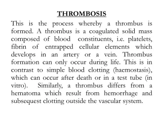
HAEMOSTASIS AND THROMBOSIS. The integrity of the circulation is maintained by blood flowing through intact vessels lined by endothelial cells. Injury to the vessel wall exposes collagen and together with tissue injury sets in motion a series of events leading to haemostasis. HAEMOSTASIS

E N D
The integrity of the circulation is maintained by blood flowing through intact vessels lined by endothelial cells. Injury to the vessel wall exposes collagen and together with tissue injury sets in motion a series of events leading to haemostasis
HAEMOSTASIS Haemostasis is a complex process depending on interactions between the vessel wall, platelets and coagulation and fibrinolytic mechanisms.
Vessel wall The vessel wall is lined by endothelium which, in normal conditions, prevents platelet adhesion and thrombus formation. This property is partly due to its negative charge but also to: ■ thrombomodulin and heparan sulphate expression ■ synthesis of prostacyclin (PGI2) and nitric oxide (NO), which cause vasodilatation and inhibit platelet aggregation ■ production of plasminogen activator Injury to vessels causes reflex vasoconstriction, while endothelial damage results in loss of antithrombotic properties, activation of platelets and coagulation and inhibition of fibrinolysis
Platelets Platelet adhesion. When the vessel wall is damaged, the platelets escaping come into contact with and adhere to collagen and von Willebrand factor that is bound below the endothelium. This is mediated through glycoprotein Ib (GPIb).
Glycoprotein IIb–IIIa is then exposed, forming a second binding site for VWF. Within seconds of adhesion to the vessel wall platelets begin to undergo a shape change, from a disc to a sphere, spread along the subendothelium and release the contents of their cytoplasmic granules, i.e. the dense bodies (containing ADP and serotonin) and the α- granules (containing platelet-derived growth factor, platelet factor 4, β-thromboglobulin, fibrinogen, VWF, fibronectin, thrombospondin and other factors). Platelet release. The release of ADP leads to a conformational change in the fibrinogen receptor, the glycoprotein IIb–IIIa complex (GPIIb–IIIa), on the surfaces of adherent platelets allowing it to bind to fibrinogen
Platelet aggregation. As fibrinogen is a dimer it can form a direct bridge between platelets and so binds platelets into activated aggregates (platelet aggregation) and further platelet release of ADP occurs. A self-perpetuating cycle of events is set up leading to formation of a platelet plug at the site of the injury
Coagulation. After platelet aggregation and release of ADP, the exposed platelet membrane phospholipids are available for the assembly of coagulation factor enzyme complexes (tenase and prothrombinase); this platelet phospholipid activity has been called platelet factor 3 (PF-3). The presence of thrombin encourages fusion of platelets, and fibrin formation reinforces the stability of the platelet plug. Central to normal platelet function is platelet prostaglandin synthesis, which is induced by platelet activation and leads to the formation of TXA2 in platelets (Fig. 8.34). Thromboxane (TXA2) is a powerful vasoconstrictor and also lowers cyclic AMP levels and initiates the platelet release reaction. Prostacyclin (PGI2) is synthesized in vascular endothelial cells and opposes the actions of TXA2. It produces vasodilatation and increases the level of cyclic AMP, preventing platelet aggregation on the normal vessel wall as well as limiting the extent of the initial platelet plug after injury
Coagulation and fibrinolysis Coagulation involves a series of enzymatic reactions leading to the conversion of soluble plasma fibrinogen to fibrin clot . Roman numerals are used for most of the factors, but I and II are referred to as fibrinogen and prothrombin respectively; III, IV and VI are redundant. The active forms are denoted by ‘a’. The coagulation factors are primarily synthesized in the liver and are either serine protease enzyme
precursors (factors XII, XI, X, IX and thrombin) or cofactors (V and VIII), except for fibrinogen, which is degraded to form fibrin.
Coagulation pathway This enzymatic amplification system was traditionally divided into ‘extrinsic’ and ‘intrinsic’ pathways. This concept is useful for the interpretation of clinical laboratory tests such as the prothrombin time (PT) and activated partial thromboplastin time (APTT) but unrepresentative and an oversimplification of in vivo coagulation. Coagulation is initiated by tissue damage. This exposes tissue factor (TF) which binds to factor VII. The TF–factor VII complex directly converts factor X to active factor Xa and some factor IX to factor IXa. In the presence of factor Xa, tissue factor pathway inhibitor (TFPI) inhibits further generation of factor Xa and factor IXa.
Following inhibition by TFPI the amount of factor Xa produced is insufficient to maintain coagulation. Further factor Xa, to allow haemostasis to progress to completion, can only be generated by the alternative factor IX/factor VIII pathway. However, enough thrombin exists at this point to activate factor VIII (and factor V) and together with factor IXa (generated by TF-factor VIIa) further activation of factor X can proceed. The presence of activated factor V dramatically enhances the conversion of prothrombin to thrombin by factor Xa. Without the amplification and consolidating action of factor VIII/factor IX, bleeding will ensue as generation of factor Xa is insufficient to sustain haemostasis.
Thrombin hydrolyses the peptide bonds of fibrinogen, releasing fibrinopeptides A and B, and allowing polymerization between fibrinogen molecules to form fibrin. At the same time, thrombin, in the presence of calcium ions, activates factor XIII, which stabilizes the fibrin clot by cross-linking adjacent fibrin molecules Factor VIII consists of a molecule with coagulant activity (VIII:C) associated with von Willebrand factor. Factor VIII increases the activity of factor IXa by ~200 000 fold. VWF functions to prevent premature factor VIII:C breakdown and locate it to areas of vascular injury. VIII:C has a molecular weight of about 350 000.
Von Willebrand Factor (VWF) is a glycoprotein with a molecular weight of about 200 000 which readily forms multimers in the circulation with molecular weights of up to 20 × 106. It is synthesized by endothelial cells and megakaryocytes and stored in platelet granules as well as the endothelial cells. The high-molecular-weight multimeric forms of VWF are the most biologically active
Physiological limitation of coagulation Without a physiological system to limit blood coagulation dangerous thrombosis could ensue. The natural anticoagulant mechanism regulates and localizes thrombosis to the site of injury.
Antithrombin. Antithrombin (AT), a member of the serine protease inhibitor (serpin) superfamily, is a potent inhibitor of coagulation. It inactivates the serine proteases by forming stable complexes with them, and its action is greatly potentiated by heparin. Activated protein C. This is generated from its vitamin K dependent precursor, Protein C, by thrombin; thrombin activation of protein C is greatly enhanced when thrombin is bound to thrombomodulin on endothelial cells. Activated protein C inactivates factor V and factor VIII, reducing further thrombin generation. Protein S. This is a cofactor for protein C, which acts by enhancing binding of activated protein C to the phospholipid surface. It circulates bound to C4b binding protein but some 30–40% remains unbound and active (free protein S).
Other inhibitors. Other natural inhibitors of coagulation include α2-macroglobulin, α1-antitrypsin and α2-antiplasmin
Fibrinolysis Fibrinolysis is a normal haemostatic response that helps to restore vessel patency after vascular damage. The principal component is the enzyme plasmin, which is generated from its inactive precursor plasminogen. This is achieved principally via tissue plasminogen activator (t-PA) released from endothelial cells. Some plasminogen activation may also be promoted by urokinase, produced in the kidneys. Other plasminogen activators (factor XII and prekallikrein) are of minor physiological importance
Plasmin is a serine protease, which breaks down fibrinogen and fibrin into fragments X, Y, D and E, collectively known as fibrin (and fibrinogen) degradation products (FDPs). D-dimer is produced when cross-linked fibrin is degraded. Its presence in the plasma indicates that the coagulation mechanism has been activated. The fibrinolytic system is activated by the presence of fibrin. Plasminogen is specifically adsorbed to fibrin and fibrinogen by lysine-binding sites. However, little plasminogen activation occurs in the absence of polymerized fibrin, as fibrin also has a specific binding site for plasminogen activators, whereas fibrinogen does not.
t-PA is inactivated by plasminogen activator inhibitor-1 (PAI-1). Activated protein C inactivates PAI-1 and therefore induces fibrinolysis. Inactivators of plasmin, such as α2-antiplasmin .and thrombin-activatable fibrinolysis inhibitor (TAFI), also contribute to the regulation of fibrinolysis.
Investigation of bleeding disorders Although the precise diagnosis of a bleeding disorder will depend on laboratory tests, much information is obtained from the history and physical examination: ■ Is there a generalized haemostatic defect? Supportive evidence for this includes bleeding from multiple sites, spontaneous bleeding, and excessive bleeding after injury. ■ Is the defect inherited or acquired? A family history of a bleeding disorder should be sought. Severe inherited defects usually become apparent in infancy, while mild inherited defects may only come to attention later in life, for example with excessive bleeding after surgery, childbirth, dental extractions or trauma. Some defects are revealed by routine coagulation screens which are performed before surgical procedures. ■ Is the bleeding suggestive of a vascular/platelet defect or a coagulation defect
Vascular/platelet bleeding is characterized by easy bruising and spontaneous bleeding from small vessels. There is often bleeding into the skin. The term purpura includes both petechiae, which are small skin haemorrhages varying from pinpoint size to a few millimetres in diameter and which do not blanch on pressure, and ecchymoses, which are larger areas of bleeding into the skin. Bleeding also occurs from mucous membranes especially the nose and mouth. Coagulation disorders are typically associated with bleeding after injury or surgery, and in more severe forms, haemarthroses and muscle haematomas. There is often a short delay between the precipitating event and overt haemorrhage or haematoma formation
Laboratory investigations ■ Blood count and film show the number and morphology of platelets and any blood disorder such as leukaemia or lymphoma. The normal range for the platelet count is 150–400 × 109/L. ■ Bleeding time measures platelet plug formation in vivo. It is determined by applying a sphygmomanometer cuff to the arm and inflating it to 40 mmHg. Two 1 mm deep, 1 cm long incisions are made in the forearm with a template. Each wound is blotted every 30 seconds and the time taken for bleeding to stop is recorded, normally between 3 and 10 minutes. Prolonged bleeding times are found in patients with platelet function defects, and there is a progressive prolongation with platelet counts less than 100 × 109/L. The bleeding time should not be performed at low platelet counts. ■ Coagulation tests are performed using blood collected into citrate, which neutralizes calcium ions and prevents clotting
The prothrombin time (PT) is measured by adding tissue factor (thromboplastin) and calcium to the patient’s plasma. The normal PT is 12–16 seconds and may be expressed as the international normalized ratio, INR The PT measures VII, X, V, prothrombin and fibrinogen (classic ‘extrinsic’ pathway) and is prolonged with abnormalities of these factors. It may also be abnormal in liver disease, or if the patient is on warfarin-
The activated partial thromboplastin time (APTT) is also sometimes known as the PTT with kaolin (PTTK). It is performed by adding a surface activator (such as kaolin), phospholipid (to mimic platelet membrane) and calcium to the patient’s plasma. The normal APTT is 30–50 seconds and depends on the exact methodology. The APTT measures XII, XI, IX, VIII, X, V, prothrombin and fibrinogen (classic ‘intrinsic’ pathway) and is prolonged with deficiencies of one or more of these factors. It is not dependent on factor VII.
The thrombin time (TT) is performed by adding thrombin to the patient’s plasma. The normal TT is 12–14 seconds, and it is prolonged with fibrinogen deficiency, qualitative defects of fibrinogen (dysfibrinogenaemia) or inhibitors such as heparin or FDPs
Correction tests can be used to differentiate prolonged times in the PT, APTT and TT due to various coagulation factor deficiencies and inhibitors of coagulation. Prolonged PT, APTT or TT due to coagulation factor deficiencies can be corrected by addition of normal plasma to the patient’s plasma. Failure to correct after addition of normal plasma is suggestive of the presence of an inhibitor of coagulation. Factor assays are used to confirm coagulation defects, especially where a single inherited disorder is suspected. Special tests of coagulation will often be required to confirm the precise haemostatic defect. Such tests include estimation of fibrinogen and FDPs, platelet function tests such as platelet aggregation and platelet granule contents.