Download
1 / 23
240 likes | 602 Views
Thrombosis. Dr Nico Lategan MBChB, MMed (Haematology). General. Coagulation/ Haemostasis: Blood clotting vs Fibrinolysis. Endothelium damage: release of TF (activates clotting) and tPA (activates fibrinolysis). Blood clotting: Virchow’s triad- vessel wall, blood flow, blood components.

E N D
Thrombosis Dr Nico Lategan MBChB, MMed (Haematology)
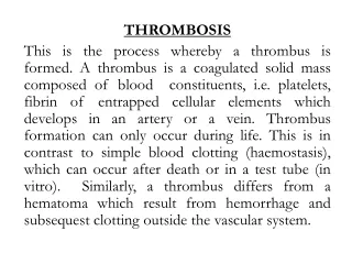
General • Coagulation/ Haemostasis: Blood clotting vs Fibrinolysis. • Endothelium damage: release of TF (activates clotting) and tPA (activates fibrinolysis). • Blood clotting: Virchow’s triad- vessel wall, blood flow, blood components. • Vessel wall: Important, especially in arterial thrombosis. • Blood flow: Stenosis, PV (RBC), CML (WCC), ET (PLT, acute leukaemia (Blasts).
Definition: Tendency to develop recurrent thrombosis at unusual sites due to enhanced thrombin generation started at a young age (<50 years).
Classification: 1. Familial – physiological 2. Non-familial (acquired) – physiological or pathological
Problems: • Confirmation of diagnosis. • Investigate, establish the cause. • Therapy: Warfarin, Heparin, Disprin. • Short term vs Long term.
Investigation: • Good history. • Family history (can be difficult). • Medication (hormones). • About recent TE. • Thorough examination. • Risk factors: previous episodes, immobilization, operations, trauma, pregnancy, obesity, younger than 40 years, homocysteinemia.
Always do basic tests: • FBC, PLT, ESR • PT, aPTT, TT, Fibrinogen
Remember: • After acute episode, it is not recommended to do a full thrombotic profile to determine the cause: Fibrinogen, F VIII, and PAI are acute phase agents. • Prot C and S, as well as AT III may be low. • Can be difficult to distinguish between liver disease, acute DIC and Warfarin therapy. • All clotting factors are produced by the liver except F VIII- probably from endothelium.
About Thrombophilia: • Usually venous TE. • Role in arterial Thrombosis? • Autosomal dominant hereditary pattern: hetero- vs homozygous inheritance. • Usually a risk factor needed in heterozygotes to be of clinical importance.
Familial: • FV-Leiden (APCR: activated prot C resistance). • Protein C (deficiency). • Protein S (deficiency). • Antithrombin III (deficiency). • Abnormal Prothrombin (PT 20210 A). • Sticky platelet syndrome.
1. FV-Leiden: • One of the most common causes for thrombophilia – 20% of clinical disease (AT, PC and PS – 5%) + risk factor. • Activated PC inhibits F Va and F VIIIa. • Inability of APC to inhibit the above complex due to mutated FV. • Heterozygous: 5-10 times increased risk for TE. • Homozygous: 50-100 times.
2. Protein C Deficiency: • Common cause (increasing TE with age). • Needs TM from endothelium wall. • Heterozygous: 50% of level of normal individuals. • Homozygous: babies are born with undetected levels (thrombi in microvascular of skin DIC necrosis purpura fulminans).
3. Protein S Deficiency: • Non-enzymatic co-factor for PC. • Binds to TM-PC. • Same properties as PC. • Two forms: free in plasma and bound to C4b binding protein (60%). Only free fraction functions as co-factor for APC. • Sometimes difficult to get accurate measures of PS because of the latter. • Like PC can be acquired: liver disease, Warfarin, pregnancy, cancer, DIC and chemo.
4. Antithrombin III Deficiency: • Common cause (incidence 1/2000 – 1/5000; heterozygotes; 50% DVT): Quantitative vs Qualitative disorder. (Acquired: DIC, cirrhosis, NS). • Bind to and inactivate thrombin, Factors IXa, Xa, XIa and XIIa (AT/heparin complex - rate of inhibition 1000-fold increased). • Not necessarily a risk factor to be involved in heterozygotes to give TE. • Increased incidence with ageing: 80% at 55 years.
5. Abnormal Prothrombin (PT 20210 A): • Common. • Increased levels of prothrombin enhanced thrombin formation. • Only way for diagnosis: DNA-PCR technique.
6. Sticky Platelet Syndrome: • Especially in arterial thrombosis (MI, TIA) and development of recurrent TE while on Warfarin. • 3 Forms. • If on aspirin, it should be stopped 14 days prior to testing. Also remember: PC, PS and AT III are inhibitors of clotting.
Non-familial (Acquired): • Antiphospholipid Syndrome: • Antibodies directed against phospholipid cell membrane = APA (Antiphospholipid Ab). • APA: ACA or LA. • Primary (PAPS) or secondary (autoimmune disorders, e.g. SLE) • ACA (Anticardiolipin Ab): IgM + IgG. • IgG: the clinically important one. • IgM: pregnancy, infection (viral), trauma and post-op. • LA (Lupus anticoagulant): Ab which affect clotting tests (LA-PTT, RVV, Kaolin). • PAPS = TE, miscarriage, IUD + ACA, LA.
Non-familial (Acquired) continued: • TPA (Tissue Plasminogen Activator): decreased levels impaired fibrinolysis. • PAI (Plasminogen Activator Inhibitor): increased levels decreased TPA. • Dysfibrinogenemia. • F XII deficiency: Hageman factor. • Fibrinogen (increased). • F VIII (increased). • Plasminogen. • Hyperhomocyteinemia – enzyme (folate).
Investigation (Thrombotic Profile): NB: Patients can be on Warfarin, but not Heparin! • FBC, PLT & ESR • PT, aPTT, TT & Fibrinogen • PC & PS • AT III • APCR (if + screening, submit for PCR) • PT 20210A (PCR) • Lupus anticoagulant (RVVT, KT, LA-PTT) • Cardiolipin antibodies (antiphospholipid syndrome) • Sticky platelet syndrome (aspirin!) • ANA screening • PNH screening
When to test: • Younger: < 50 years, recurrent TE, unusual sites, TE on Warfarin. • Not ideal to test after acute episode (inhibitors of clotting may be low). • Ideal: test after 6 weeks after settlement of hemostasis. • Most patients are on Warfarin then (PC & PS are Vit K dependent, may be falsely low). • My view: if long-term Warfarin is planned, do immediately/ according to duration of treatment it can be done after cessation of treatment.
When to test (continued): • Practical (my experience): before treatment – if AT, PS, PC are low repeat after Rx has been stopped. • SPS: platelet aggregation studies (problem: sometimes aspirin cannot be stopped). • Remember the effect of the vessel wall on clotting, especially in arterial thrombosis. • Every woman on contraception, HRT?
Treatment: • Heparin: unfractioned vs LMW. • Heparin – PTT. LMW – anti FXa activity. • NB: LMW does not affect PTT. • Warfarin: venous, antiphospholipid syndrome. • Warfarin: PT / INR (not % due to lab variation). • Aspirin: arterial – SPS. • Individualized patients: ex single episode of thrombosis in patient with FV-Leiden post-op lifelong treatment unnecessary (short to medium term). • Lifelong: recurrent episodes, episodes on Warfarin, ? spontaneous episode with proven cause (DVT vs PE).
Duration of Treatment: • 6 weeks • 3 months • 6 months INR Range: • Single episode: 2-3 • Recurrent episode: 3-3.5 • PE: 2-3/ 3-3.5