Download
1 / 35
390 likes | 788 Views
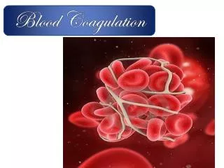
NORMAL HAEMOSTASIS:. It is a complex process depending on interaction between vessel wall, platelets and coagulation factors. NORMAL HAEMOSTASIS. Vessel wall.

E N D
NORMAL HAEMOSTASIS: It is a complex process depending on interaction between vessel wall, platelets and coagulation factors.
NORMAL HAEMOSTASIS Vessel wall Injury to vessel leads to immediate reflex vasoconstriction controlling hemorrhage, at the same time damage of endothelium results in activation of platelets and coagulation release of serotonin and thromboxan A2 from activated platelets contribute to vasoconstriction. Platelets Coagulation & fibrinolysis
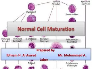
NORMAL HAEMOSTASIS They are produced in B.M from megakaryocytes under the influence of thrombopoetin which inhance production, maturation and release. Vessel wall Platelets Coagulatio & fibrinolysis
NORMAL HAEMOSTASIS Structure of platelets: They are disc shaped with 2-3 microns diameters with blue and granular core. On electron microscope it has 2 Zones: • 1- Peripheral zone: made up from protein, lipids and CHO. • A-platelet membrane:Containing various cell receptors and enzymes. • B-system of microtubules and microfilaments:Forming a channel where factors secreted by platelet reaching surrounding media. • 2- Central zone: Containing various organelles formed of granules, vacules and dense particles. Granules are glycogen, which is the main source of energy for platelets metabolism. Dense particles contain enzymes, serotonin and ADP, which are released from platelets during platelet secretion. Platelets
NORMAL HAEMOSTASIS Platelets functions: • 1- Platelets adhesion: To collagen which depends on platelet membrane receptors, glycoproteins Ia (GP-Ia) which binds directly to collagen and (GP-Ib) which binds to Von-Willebrand factor (VWF) in plasma and VWF in turn adheres to collagen following adhesion shape change from disc to sphere release of content of platelets granules (platelet release): • ADP & serotonin and TXA2 • Platelet derived growth factor. • Platelet factor 4. Platelets
NORMAL HAEMOSTASIS Platelets functions: 2- Platelets aggregations: ADP leads binding of fibrinogen to platelet platelet aggregation forming plug at site of injury. Also of normal platelet function is platelet prostaglandin synthesis which is induced by platelet activation and leads to formation of TXA2 powerful vasoconstrictor
NORMAL HAEMOSTASIS Platelets functions: 3-Platelet secretion : As a result of platelet aggragation; platelet factor-3 is released which intiates blood coagulation ground platelets , thrombin encourage fusion of platelet and fibrin formation reinforce stability platelet plug . 4- Platelet contraction:Finally, clot retraction occurs by contraction of protein thromasthenin which together with fibrin around form hemostatic plug. Prostacyclin (PGI2) is synthesized in vascular endothelium and oppose the action of TXA2 and it increase level of cyclic AMP preventing platelet aggregation on normal vessel wall as well as limiting the extent of platelet plug after injury.
NORMAL HAEMOSTASIS Vessel wall • Coagulation cascade involves a series of enzymatic reactions leading to conversion of soluble plasma fibrinogen to fibrin clot. • Coagulation factors are synthesized in the liver. Platelets Coagulation & fibrinolysis
NORMAL HAEMOSTASIS Coagulation pathways (A) Extrinsic pathway. Coagulation is initiated by tissue factor, which is expressed on the surface of endothelial cells coming into contact with plasma after injury. Complex of activated factor VII (TF:VIIa) Xa. (B) Intrinsic pathway Factor IX is activated by complex of tissue factor and factor VII. IXa together with factor VIII and calcium ion activate factor X. Xa conversion of prothrombin to thrombin.
NORMAL HAEMOSTASIS Coagulation pathways (C) Common pathway Fibrinogen fibrinpeptide A&B and Polymerization of fibrinogen Fibrin. At the same time thrombin in presence of Ca ion activate factor VIII, which stabilize fibrin clot by cross-linking. Factor VIII is associated with Von willebrand factor whose function is to stabilized VIII and to promote platelet endothelial interactions.
NORMAL HAEMOSTASIS: LIMITATION OF COAGULATION: • Coagulation is limited by: • Removal of activated coagulation factors by rapid blood flow. • Plasma inhibitors of activated coagulation factors. • Fibrinolysis • Inhibition of coagulation: • Antithrombin: is potent inhibition of coagulation. Its action is potentiated by heparin. • Activated protein C:This generated from its Vit K dependent precursor. It is activated when thrombin is bound to thrombomodulin (an endothelial receptors), activated protein C destroy factor V and VIII. • Protein S:This co-factor for protein c allowing binding of protein c to platelet surface. • Other inhibitors: -α2 Macroglobulin.-α1 antiplasmin.-α1antitrypsin.-Heparin Cofactor II.
NORMAL HAEMOSTASIS: FIBRINOLYSIS • It helps to restore vessel potency by dissolution of fibrin. • Normal plasma protein plasminogen (formed in the liver) is converted by plasminogen activators derived from plasma endothelial cells, platelets, leukocytes and urine into plasmin.
NORMAL HAEMOSTASIS: FIBRINOLYSIS • 2 types of plasminogen activator: • Tissue plasminogen activator formed in the vascular endothelium (t-PA). • Tissue plasminogen activator formed in the kidney (Urokinase). • t-PA is inactivated by plasminogen activator inhibitor I. All of which regulate fibrinolysis. • Plasmin (serine protease) breaks fibrinogen and fibrin into fragments X, Y, D and E degradation of cross linked fibrin yield D-dimer and D-dimer E fragment. • All fibrinogen/fibrin degeneration products (FDPs) are normally rapidly cleared from circulation by the liver and R.E.S.
NORMAL HAEMOSTASIS: INVESTIGATION OF HEMOSTASIS 1. Tests of vascular integrity: • Also affected by thrombocytopenia or thrombocytopathy. • Bleeding time (N: 6-10 min). • Hess’s test ( Capillary resistance test): Area 5 Cm in diameter marked on antecubital fossa and sphygmomanometer is inflated to 80 mm Hg for 5 minutes. Normally 3-5 purpuric eruptions appear. • 2. Test for platelets: • Blood count and film: Show number and morphology of platelets & any blood disorder as leukemia. • Direct test of platelet function: • Platelet aggregation: By adding of aggregating agents as ADP, collagen, thrombin, adrenaline and antibiotic Ristocetin. • Platelet adhesion: (to glass surface): Decrease in Von willebrand disease, uremia and thrombocytopenia & increase in arterial and venous disease and after surgery. • Platelet antibodies: Its presence indicates autoimmune thrombocytopenia.
NORMAL HAEMOSTASIS: INVESTIGATION OF HEMOSTASIS 3.Tests for blood coagulation (1) Prothrombin time (PT) (N: 16-18 sec). It is prolonged with abnormality of factor VII, X, V or prothrombin and fibrinogen (extrinsic system). It is used for control of anticoagulant therapy. (2) Activated partial thromboplastin time (APTT) (N: 30-50 sec). It is prolonged with deficiencies or inhibitors of I, II, V, VIII, IX, X, XI, XII. (Intrinsic and common pathway). (3) Thrombin time (TT) (N: 10-20 sec) : It is prolonged with fibrinogen deficiency and dysfibrinogenemia or inhibitors as heparin or FDPs. (4) Plasma fibrinogen level (N: 150-400 mg%) Increase in pregnancy & postoperative. Decrease in DIC & congenital hypofibrinogenemia. (5) Coagulation factor assay: Value below 40% is abnormal. (6) Whole blood clotting time (N: 5-10 min): Normal test excludes cross deficiency in the coagulation time but neglect minor changes . (7) Fibrinogen/fibrin degradation products (FDPs) (N: 10 mcg/ml): It is increase in all cases of DIC and in thromboembolic disease . Very high level above 600 mg/ml occur in severe DIC.
NORMAL HAEMOSTASIS: INVESTIGATION OF HEMOSTASIS
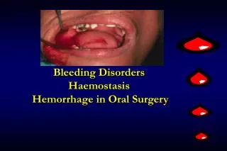
HAEMORRHAGIC DISORDERS HAEMORRHAGIC DISORDERS • Vascular defect. • Platelet defect. • Coagulopathy
HAEMORRHAGIC DISORDERS VASCULAR DISORDERS NON THROMBOCYTOPENIC PURPURA Characterized by easy bruising and bleeding into the skin and sometimes from mucus membrane. Both bleeding time & platelet count are normal.
HAEMORRHAGIC DISORDERS VASCULAR DISORDERS NON THROMBOCYTOPENIC PURPURA • I. Congenital: • Hereditary hemorrhagic telangiectesia. • Dilatation of capillary& arterioles in skin & mucus membrane present with epistaxis, hemoptysis and with GIT bleeding. • Connective tissue disorders ( Ehler’s Danlos syndrome). • II. Acquired: • 1- Infection : • Severe infection caused by damage to vascular endothelium, septicemia. • Meningococal infection. • Typhoid. • Measels. • 2. Allergic: • Henoch-schonlein purpura. • 3. Autoimmune disorders (SLE, Rheumatoid disorders) • Due to vasculitis papules & elevated • 4. Drugs: • Steroids *Sulphonamides • 5. Others: • Senile purpura • Due to atrophy of vascular supporting tissues. • Scurvy. • Easy bruising syndrome.
HAEMORRHAGIC DISORDERS VASCULAR DISORDERS HENOCH-SCHONLEIN PURPURA Affect children following streptococal sore thorat. 1- Purpura: There is capillaritis and arteriolitis associated with deposation of IgA and characterized by: purpura over the extensor surfaces of arm, leg and buttocks which may be itchy. 2- Arthritis. : tender swelling of the jiont with periarticular affection 3- Abdominal pain: due to affection of submucosal intestinal blood vesels. 4- Glomerulonephritis which is usually benign, only 9-10 pass to chronic TREATMENT: Corticosteroid
HAEMORRHAGIC DISORDERS PLATELET DISORDERS • Normal platelet count 150.000 – 400.000/Cmm. • Platelets are produced in bone marrow from megakaryocytes under influence of thrombopoietin which enhance production, maturation and release of blood platelets
HAEMORRHAGIC DISORDERS PLATELET DISORDERS CLASSIFICTION OF PLATELETS DISORDERS: • Thrombocytopenia. • Thrombocythemia. • Thrombocytpathies. (functional disorders)
HAEMORRHAGIC DISORDERS PLATELET DISORDERS CAUSES OF THROMBOCYTOPENIA: • 1. Impaired production • Marrow aplasia (primary or sechondry). • Marrow infeltiration (Leukemia, lymphoma, carcinoma and myelofibrosis. • Drugs e.g : Co-trmexazole, Thiazide, Cyloxic, ethanol. • 2. Excessive distruction • [A] Immune: • Autoimmune: (Idiopathoc Thrombocytopenic purpura) • Sechondry immune: (SLE, CLL, viral infection, drugs e.g: asprin, sulpha, thiazide, quinine, paracetamol, barbiturate). • Alloimmune : post transfusion purpura. • [B] Mechanical : • prothetic heart valve. • Thrombotic thrombocytopenic purpura. • 3. Increased platelet consumption • DIC. • Giant hemangioma in children. • 4. Increased platelet sequestration • Hypersplenism. • 5. Increased platlet dilution • Massive transfusion of stored blood.
HAEMORRHAGIC DISORDERS IDIOPATHOC THROMBOCYTOPENIC PURPURA (ITP) It is due to immune destruction of plateles. The sensetized platelets are removed by R.E.S. due to IgG antibody. The antibody coated platelet are removed by spleen. There are 2 types: 1- Acute : Seen in children but may occur in adult often following viral infection due to deposition immune complexes on platelets, it is usually selflimited. 2- Chronic: Is usually seen in adult women either idiopathic or in association with other autoimmune disorder as SLE, thyroid disease or with autoimmune hemolytic anemia ( Evan’s syndrome), CLL after viral infection. It usually occurs in intermittent form
HAEMORRHAGIC DISORDERS IDIOPATHOC THROMBOCYTOPENIC PURPURA (ITP) • CLINICAL FEATURES • Major Hemorrhage is rare as is seen only in patients with sever thrombocytopenia below 10,000/Cmm. • Bleeding occurs either spontaneous or after truma in the form of petechiae or purpuric eruption easy brusing, epistaxis and menorrhagia, GIT or genitourinary bleeding. • Splenomegaly is rare.
HAEMORRHAGIC DISORDERS IDIOPATHOC THROMBOCYTOPENIC PURPURA (ITP) • DIAGNOSIS: 3 negative finding • Absence of fever. • Absence of lymphadenopathy or marked splenomegally(10% just palpable) • Normal ESR & serology
HAEMORRHAGIC DISORDERS IDIOPATHOC THROMBOCYTOPENIC PURPURA (ITP) INVESTIGATION: 1- C.B.C - Decrease platlets count. - Normal RBCs and WBCs. - Rarely +Ve coomb’s test. 2- B. M - Normal granulopoiesis and erythropoiesis. -Abundant megakaryocytes with no excessive budding due to rapid release into circulation. 3-Platelets antibodies: not essential for confirmation of diagnosis.
HAEMORRHAGIC DISORDERS IDIOPATHOC THROMBOCYTOPENIC PURPURA (ITP) D. D: - Drug reaction. - SLE. - aplastic anemia. - Leukemia. – B.M infilteration. Prognosis: Spontaneous remission & relapse. Cerebral hemorrhage may occur in early months.
HAEMORRHAGIC DISORDERS IDIOPATHOC THROMBOCYTOPENIC PURPURA (ITP) TREATMENT: Acute: treatment required only when count is below 20.000/Cmm or there is bleeding. Chronic: spontaneous remission is rare. Aim: (1) reduce production of autoantibodies. (2) reduce removal of antibody coated platelets.
HAEMORRHAGIC DISORDERS IDIOPATHOC THROMBOCYTOPENIC PURPURA (ITP) TREATMENT: 1- Corticosteroids: Corner stone of treatment. - It diminish the antibodies formation. - Suppress phagocytosis of antibody coated platelets. 40 – 60 mg prednisone / day for 2-4 weeks, after platelet becomes normal it is tapered over 4 – 8 weeks. Our targe is platelet above 60.000/Cmm. 2- Splenectomy: For patient not responding to corticosteroids. 80% of patients respond although 20% remain thrombocytopenic (N.B: steroid & platelet transfusion are used to support patient in pre & post operative periods).
HAEMORRHAGIC DISORDERS IDIOPATHOC THROMBOCYTOPENIC PURPURA (ITP) TREATMENT: 3- Intravenous infusion of high dose immunoglobulin. Produce rapid rise due to blockade of Fc receptor on macrophage in the spleen. The effect is transient but is indicated in acute hemorrhage and in preparing patient with chronic ITP for surgery . 4- Immunosuppresive drugs: Vincristine, cyclophosphamide and azathioprine may be used in patients not responding to corticosteroids or splenectomy. 5- Anabolic steroids: Danazole 200 mg t.d.s. 6- Platelet transfusion: In severe bleeding or with emergency surgery, it should be transfused within 6 hs of their collection.
HAEMORRHAGIC DISORDERS OTHER IMMUNE THROMPOCYTOPENIA: 1-Drugs: By forming drug antibody immune complex or antigenic drug platelet complex e.g: quinine, pencilline or methyl dopa. 2-Post-Transfusion Purpura: 2-12 days after blood transfusion associated with platelet alloantibodies.
HAEMORRHAGIC DISORDERS FUNCTIONAL PLATELET DISORDER ( thrombocytopathies) • These are usually associated with excessive brusing, purpura, bleeding from mucus membrane. • The platelet count is normal and bleeding time is prolonged and abnormal platelet function ( platelet adhesion, aggregation, platelet release and platelet factor III avaliablity).
HAEMORRHAGIC DISORDERS FUNCTIONAL PLATELET DISORDER ( thrombocytopathies) (1) Inherited type: Glanzman’s disease: There is deficiency of platelet membrane glycoprotein IIb/IIIa, platelet fail to adhere to glass or aggregate with addision of aggregating agent (ADP, Adrenaline, Collagen and Ristocetin). Brenard Soalier disease: Due to lack of platelet membrane glycoprotein Ib/IX the binding site for vWF. Characterized by giant platelets. Platelet aggregation is normal to all except for Ristocetin. Storage-pool disease: The abnormality in platelet release of ADP charcterized by decrese agregregation only by collagen.
HAEMORRHAGIC DISORDERS FUNCTIONAL PLATELET DISORDER ( thrombocytopathies) (2) Acquired Thrombocytopathies: • 1- Drugs: • Aspirin ( cyclo-oxygenase inhibitor). • Non-steroidal anti inflammatory (prostaglandin synthetase inhibitors). • Dipyridamol. • They impair function of platelet and accentuate bleeding. • 2- Uremia and liver diseases. • 3- Paraprotenemia. • 4- Myeloproliferative disorder. • TREATMENT • Offending drug should be stopped. • Severe bleeding need platelet transfusion. • Desmopressin (DD,AVP) may be helpful.