Download
1 / 40
440 likes | 534 Views
Reflexes. Definition. A reflex may be defined as an immediate and involuntary response to a stimulus. A reflex is a fast response to a change in the body's internal or external environment in an attempt to restore homeostasis. Reflexes and diagnosis.

E N D
Definition • A reflex may be defined as an immediate and involuntary response to a stimulus. • A reflex is a fast response to a change in the body's internal or external environment in an attempt to restore homeostasis.
Reflexes and diagnosis • Evaluation of a reflex can aid a doctor in diagnosing a problem. • A reflex which stops functioning or functions abnormally may indicate that a particular central or peripheral conduction pathway in the body has been damaged.
Muscle reflexes • Muscle reflexes help determine how responsive the spinal cord is. • It may become so sensitive that just tapping the tendon of the knee with the tip of your finger can cause the leg to jump a considerable distance.
Evaluation of neurological impairment • It is part of neurological examination. • You can evaluate neurological impairment by testing reflexes using a stopwatch to time the reflex response.
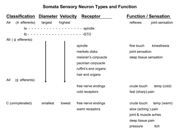
Mechanism of deep reflexes • When a skeletal muscle with an intact nerve supply is stretched, it contracts. This response is called the stretch reflex. • The stimulus that initiates the reflex is stretch of the muscle, and the response is contraction of the muscle being stretched. • The sense organ is the muscle spindle. The impulses originating in the spindle are conducted in the CNS by fast sensory fibers that pass directly to the motor neurons which supply the same muscle.
How knee jerk happens • The tapping of the tendon applies a stretch to quadriceps tendon and muscle spindles. • The sudden stretch of the patellar tendon stimulate the muscle spindle and increase their charge to the spinal cord. • Some of these messages continue up to higher centers in the brain, and some to the motor neuron of the muscle which cause muscle to contract.
Reciprocal inhibition • Interneurons or interconnecting neurons sends inhibitory messages to the motor neurons. • This maintain proper balance for muscle activity for postural control this is called Reciprocal inhibition.
Inverse stretch reflex • when the tension becomes great enough, contraction suddenly ceases and the muscle relaxes. This relaxation in response to strong stretch is called the inverse stretch reflex or autogenic inhibition. • The receptor for the inverse stretch reflex is in the Golgi tendon organ.
DEEP TENDON REFLEXES • persuade the patient to relax. • position the limbs properly and symmetrically. • Strike the tendon briskly using a rapid wrist movement. • Your strike should be quick and direct • You may use either the pointed or the flat end of the hammer.
Grading of reflexes 4+ Very brisk, hyperactive, with clonus (rhythmic oscillations between flexion and extension) 3+ Brisker than average; possibly but not necessarily indicative of disease 2+ Average; normal 1+ Somewhat diminished; low normal 0 No response
Abnormalities Hyperactive reflexes suggest central nervous system disease. Sustained clonus confirm it. Reflexes may be diminished or absent when sensation is lost, when the relevant spinal segments are damaged, or when the peripheral nerves are damaged. Diseases of muscles and neuromuscular junctions may also decrease reflexes.
Biceps Reflex (C5, C6) The patient’s arm should be partially flexed at the elbow with palm down. Place your thumb or finger firmly on the biceps tendon. Strike with the reflex hammer so that the blow is aimed directly through your digit toward the biceps tendon. observe flexion at the elbow, and watch for and feel the contraction of the biceps muscle.
The Triceps Reflex (C6, C7) Flex the patient’s arm at the elbow, with palm toward the body, and pull it slightly across the chest. Strike the triceps tendon above the elbow. Use a direct blow from directly behind it. Watch for contraction of the triceps muscle and extension at the elbow.
If you have difficulty getting the patient to relax, try supporting the upper arm. Ask the patient to let the arm go limp, as if it were “hung up to dry.” Then strike the triceps tendon.
(Brachioradialis Reflex (C5, C6 • The patient’s hand should rest on the abdomen or the lap, with the forearm partly pronated. • Strike the radius about 1 to 2 inches above the wrist. • Watch for flexion and supination of the forearm.
Knee Reflex (L2,L3,L4) • The patient may be either sitting or lying down as long as the knee flexed. • Briskly tap the patellar tendon just below the patella. • Note contraction of the quadriceps with extension at knee. • A hand on the patient’s anterior thigh lets you feel this reflex.
Two methods are useful in examining the supine patient. Supporting both knees at once, as shown. Sometimes you may wish to rest your supporting arm under the patient’s opposite leg. Some patients find it easier to relax with this method.
The Ankle Reflex (primarily S1) If the patient is sitting, dorsiflex the foot at the ankle. Persuade the patient to relax. Strike the Achilles tendon. Watch and feel for plantar flexion at the ankle.
When the patient is lying down, flex one leg at both hip and knee and rotate it externally so that the lower leg rests across the opposite shin. Then dorsiflex the foot at the ankle and strike the Achilles tendon.
SUPERFICIAL REFLEXES • This group of reflexes includes the plantar response, the superficial abdominal reflex and cremasteric reflex. • These are polysynaptic reflexes, which are evoked by cutaneous stimulation.
The Planter Response (L5, S1) • With an object such as a key or the wooden end of an applicator stick, stroke the lateral aspect of the sole from the heel to the ball of the foot, curving medially across the ball. • Use the lightest stimulus that will provoke a response, but be increasingly firm necessary. • Note movement of the toes, normally flexion.
Dorsiflexion of the big toe, often accompanied by fanning of the other toes, constitutes a Babinski response It often indicates a central nervous system lesion in the corticospinal tract.(pyramids)
Notes Babinski response is normal in infants till walking. A Babinski response may be seen in unconscious states due to drug or alcohol intoxication or in the postictal period following a seizure. A marked Babinski response is occasionally accompanied by reflex flexion at hip and knee. Some patients withdraw from this stimulus by flexing the hip and the knee. Hold the ankle, if necessary, to complete your observation. It is sometimes difficult to distinguish withdrawal from a Babinski response.
The Abdominal Reflexes Test the abdominal reflexes by lightly but briskly stroking each side of the abdomen, above (T8, T9, T10) and below (T10, T1l, T12) the umbilicus, in the directions illustrated. Use a key,wooden end of a cotton-tipped applicator, or a tongue blade twisted and split longitudinally. Note the contraction of the abdominal muscles and deviation of the umbilicus toward the stimulus.
Abdominal reflexes may be absent in both central and peripheral nervous system disorders
Clonus If the reflexes seem hyperactive, test for ankle clonus. Support the knee in a partly flexed position. With your other hand, dorsilflex and plantar flex the foot a few times while encouraging the patient to relax, and then sharply dorsiflex the foot and maintain it in dorsiflexion. Look and feel for rhythmic oscillations between dorsiflexion and plantar flexion. In most normal people, the ankle does not react to this stimulus. A few clonic beats may be seen and felt, especially when the patient is tense or has exercised.
Notes Sustained clonus indicates central nervous system disease. The ankle planter flexes and dorsiflexes repetitively and rhythmically.