Download
1 / 49
760 likes | 1.71k Views
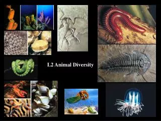
Animal Diversity. Figure 32.4 A traditional view of animal diversity based on body-plan grades. Figure 32.8 Animal phylogeny based on sequencing of SSU-rRNA. Figure 32.5 Body symmetry. Figure 32.13x Burgess Shale fossils.

E N D
Figure 32.4 A traditional view of animal diversity based on body-plan grades
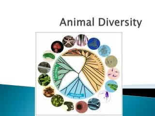
Figure 32.8 Animal phylogeny based on sequencing of SSU-rRNA
Figure 32.13 A sample of some of the animals that evolved during the Cambrian explosion
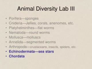
Phylum Porifera • Sponges • “colony” of flagellated cells • individual cells can potentially regenerate into a new individual
Phylum Cnidaria • Hydras, jellyfish, sea anemones, corals • gastrovascular cavity • stinging cells • Radiata
Phylum Ctenophora • Comb jellies • comblike ciliary plates • gastrovascular cavity • Radiata
Phylum Platyhelminthes • Flatworms • dorsoventrally flattened • no segmentation • gastrovascular cavity • bilateral, no coelom, protostome
Phylum Rotifera • Ciliated crown • no digestive system • bilateral, pseudocoelomates, protostome
Phylum Nematoda • Roundworms • unsegmented • no circulatory system • bilateral, pseudocoelomate, protostome
Lophophorates - several phyla • Bryozoans, lampshells (brachiopods) • bilateral, coelomate, protostome
Figure 33.14 Lophophorates: Bryozoan (left), brachiopod (right)
Phylum Mollusca • Clams, snails, squids • foot, visceral mass, mantle • bilateral, coelomate, protostome
Phylum Annelida • Segmented worms • bilateral, coelomate, protostome
Phylum Arthropoda • Crustaceans, insects, spiders • segmented body, jointed appendages, exoskeleton • bilateral, coelomate, protostome
Figure 32.7 A comparison of early development in protostomes and deuterostomes
Phylum Echinodermata • Starfish, sea urchins • bilateral, coelomate, deuterostome
Phylum Chordata • Lancelets, tunicates, vertebrates • notochord, nerve cord • bilateral, coelomate, deuterostome