Download
1 / 24
260 likes | 342 Views
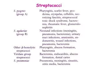
Explore the characteristics, classification, pathogenesis, enzymes, toxins and diseases caused by Streptococci, focusing on Streptococcus pyogenes.

E N D
Streptococci Part 1 Dr. Salma
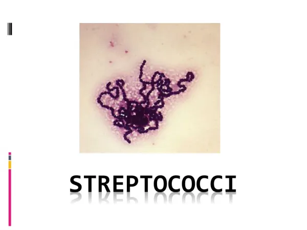
Morphology and Identification Gram-positive cocci arranged in chains or pairs. Most group A, B, and C strains produce capsules. Most strains grow as discoid colonies, 1-2 mm in diameter. Catalase-negative. Grow better in media enriched with blood or tissue fluid. Most are facultative anaerobic and some are capnophilic. For most species growth and hemolysis are aided by incubation in 10% CO2.
Staphylococcus Streptococcus
Classification Hemolysis a-hemolysis: incomplete lysis of RBC with the formation of green pigment. b-hemolysis: complete hemolysis No hemolysis Lancefield classification: a serologic classification (A to V) Biochemical reactions are used for species that can not be classified into the Lancefield classification (nongroupable), e.g. viridans streptococci.
Streptococcus pyogenes Capsule: antiphagocytosis.The capsule of group A streptococci is composed of hyaluronic acid. Group-specific cell wall antigen (Lancefield group A) Carbohydrate A dimer of N-acetylglucosamine and rhamnose. M protein T protein: type-specific; function unknown. M-like proteins: binds IgM, IgG and a2-macroglobulin; interfere with phagocytosis. Lipoteichoic acid: binds to epithelial cells. Protein F: a major adhesin of S. pyogenes, binding with fibronectin.
Streptococcus pyogenes Pathogenesis(via invasiveness and production of toxins) Adherence to the epithelial cells; >10 adhesion molecules invasion into the epithelial cells; mediated by M protein and protein F important for persistent infections and invasion into deep tissues avoiding opsonization and phagocytosis; M protein, M-like proteins, and C5a peptidase producing enzymes and toxins
Streptococcus pyogenes Enzymes and toxins Streptokinase(fibrinolysin) Can lyse blood clots and may be responsible for the rapid spread of the organism. Used (IV injection) for treatment of pulmonary emboli, coronary artery thrombosis and venousthrombosis. Streptodornase(DNases A to D) Decreases viscosity of DNA suspension. A mixture of this and streptokinase is used in enzymatic debridement-liquifies exudates and facilitates removal of pus and necrotic tissue. Hyaluronidase(spreading factor): Destroys connective tissue and aids in spreading infecting bacteria. C5a peptidase Prevents streptococci from C5a-mediated recruitment and activation of phagocytes, and is important for survival of S. pyogenes in tissue and blood.
Streptococcal pyrogenic exotoxins (Spe) Produced by both the scarlet fever strains and new invasive S. pyogenes strains. More than four serologically distinct toxins (SpeA, B, C and F). They are superantigens (except for SpeB, which is a cysteine protease)and may exhibit the followingbiological activities: Enhances release of proinflammatory cytokines (pyrogenicity) causes skin rash Immunosuppression Spe is associated with streptococcal toxic shock syndrome or other invasive S. pyogenes diseases.
Hemolysins Streptolysin O: O2-labile; causes hemolysis deep in blood agar plates. ASO (antistreptolysin O) titer >160-200 units suggests recent infection or exaggerated immune response to an earlier respiratory infection. However, skin infection does not induce ASO. Streptolysin S: O2-stable. Causes b-hemolysis on the surface of blood agar plates.Cell-bound, not antigenic. Produced in the presence of serum. Kills phagocytes by releasing the lysosomal contents after engulfment.
Streptococcus pyogenes Epidemiology S. pyogenes can transiently colonize the oropharynx and skin. Diseases are caused by recently acquired strains that can establish an infection of the pharynx or skin. S. pyogenes causes pharyngitis mainly in children of 5 to 15 years old. The pathogen is spread mainly by respiratory droplets. Crowding increases the opportunity for the pathogen to spread, particularly during the winter months. Soft tissue infections are preceded by skin colonization and the organisms are introduced into the superficial or deep tissue through a break in the skin.
Streptococcus pyogenes Clinical Diseases 1. Local infection with S. pyogenes Streptococcal sore throat (pharyngitis), and scarlet fever. Streptococcal pyoderma (impetigo, local infection of superficial layers of skin). Strains that cause skin infections are different from those that cause pharyngitis.
2. Invasion by S. pyogenes Invasion from respiratory tract: otitis media, sinusitis, pneumonia, meningitis, osteomyelitis, and arthritis. Invasion from skin: erysipelas, cellulitis, and necrotizing fasciitis. Diffuse and rapidly spreading infection that extends along lymphatic pathways with only minimal local suppuration. Sepsis (streptococcal toxic shock syndrome, STSS): the organism is introduced into the subcutaneous tissue through a break in the skin cellulitis necrotizing fasciitis systemic toxicity, multiple organ failure, and death (mortality > 40%).
3. Poststreptococcal diseases (occurs 1-4 weeks after acute S. pyogenes infection, hypersensitivity responses) Rheumatic fever: most commonly preceded by infection of the respiratory tract. Inflammation of heart (pancarditis), joints, blood vessels, and subcutaneous tissue. Results from cross reactivity of anti-M protein Ab and the human heart tissue. Acute glomerulonephritis: preceded by infection of the skin (more commonly) or the respiratory tract. Symptoms: edema, hypertension, hematuria, and proteinuria. Initiated by Ag-Ab complexes on the glomerular basement membrane. * Rheumatic fever can be reactivated by recurrent streptococcal infections, whereas nephritis does not.
S. agalactiae(group B, b-hemolytic, contains type-specific capsular polysaccharides which is the most important virulence factor and can induce protective antibodies; may colonize at lower gastrointestinal tract and genitourinary tract) Neonatal sepsis or meningitis Early-onset (during the first week of life): infection acquired in utero or at birth. Pneumonia is common in addition to meningitis. Late-onset (older infants): infection acquired from an exogenous source. (Premature infants are at greater risk.) Infection of pregnant women Urinary tract infections, amnionitis, endometritis, and wound infections Infection in men and nonpregnant women Patients are generally older and have underlying conditions. Bacteremia, pneumonia, bone and joint infections, skin and soft tissue infections. Mortality is higher.
Viridans streptococci(a-hemolytic or nonhemolytic, most are nongroupable; they, except for S. suis, are divided into 5 subgroups based on the specific diseases they cause) These streptococci colonize the oropharynx, GI tract, and GU tract; rarely on the skin surface. Diseases: Subacute endocarditis(group: Mitis) Intra-abdominal infections(group: Anginosus) Dental caries (group: Mutans) Cariogenicity of S. mutans is related to its ability to synthesize glucan from fermentable carbohydrates (e.g. sucrose) as well as to modify glucan in promoting increased adhesiveness.