Download
1 / 46
460 likes | 518 Views
Reproduction a Dr. Production. Does a 5 year old boy have mitotic divisions occurring? Does a 5 year old boy have meiotic divisions occurring? Does a 5 year old girl have mitotic divisions occurring? Does a 5 year old girl have meiotic divisions occurring? Mitosis vs Meiosis.

E N D
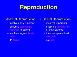
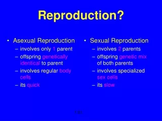
Does a 5 year old boy have mitotic divisions occurring? • Does a 5 year old boy have meiotic divisions occurring? • Does a 5 year old girl have mitotic divisions occurring? • Does a 5 year old girl have meiotic divisions occurring? • Mitosis vs Meiosis
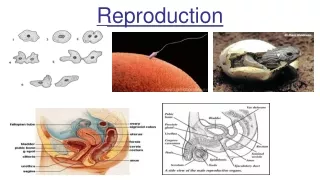
Purpose of Male Reproductive System • Spermatogenesis • Propel sperm to egg
And now located in a side view of a more complete male anatomy:
The testes, within the scrotum, both respond to and manufacture hormones. FSH from the anterior pituitary stimulates spermatocytes in the seminiferous tubules to undergo meiosis and maturation to become active sperm.
Testis • Each testicular lobule contains several coiled seminiferous tubules (ST), site of sperm production • Each ST ~ 1.3 ft in humans. Total length of ST almost the length of a football field
LH from the anterior pituitary stimulates interstitial cells, clusters of cells wedged in among the seminiferous tubules, to produce and secrete androgens (male hormones), particularly testosterone.
Sperm that are nearly mature are at the center (the lumen) of seminiferous tubules. They are collected from individual tubules into the epididymus, where they complete maturation. Passage takes about 20 days.
They are pushed from the epididymus into the vas deferens, which is the duct carrying them to the rear end of the penis, at the time of ejaculation. Before entering the penis to be ejaculated, three other sets of glands contribute fluids to what will be semen.
The seminal vesicles secrete a fluid (60% of semen) that contains vitamins and fructose to nourish sperm. The fluid also contains prostaglandins, hormones that are believed to cause contractions in the female reproductive tract to help get sperm to their ‘target’.
The prostate gland makes an opaque whitish alkaline fluid that helps protect sperm from any residual urine in the male tract and potential problems with pH in the female tract. 20% of semen.
The bulbo-urethral glands (called Cowper’s gland) secrete a clear mucous alkaline lubricant, which sometimes makes up pre-ejaculatory fluid, 10% of semen.
Ejaculation is a carefully controlled event. It occurs in at least two steps: 1) as sexual arousal peaks, muscles in the epididymus, seminal vesicles, vas deferens, and prostate all contract. As well, two sphincters associated with the prostate contract. One blocks passage of urine into the urethra. The other, further ‘downstream’, blocks flow of semen. 2) In phase 2 the second sphincter relaxes, and muscular contractions in the base of the penis moves the semen through the urethra and ejects it. Packing semen tightly between the sphincters then propelling it makes for forceful ejaculation.
The male penis is erectile, and so are the female labia (majora and minora), clitoris, and, in case you aren’t aware, also the lips and parts of the nose in both sexes. The most densely innervated and sensitive parts in both males and females are their respective glans and prepuces. In males, the prepuce is frequently surgically removed (called circumcision).
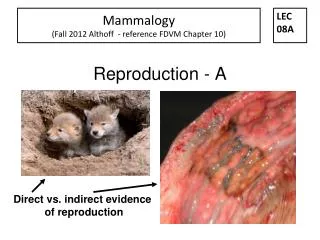
Purpose of Female Reproductive System • Oogenesis • Nurture egg
The ovary is the female gonad. In it oogonia (cells that will undergo meiosis) are randomly chosen (usually 1 per month) to undergo meiosis and develop into mature eggs.
The ovary (about the size of an almond) has a bumpy surface. Each bump is a follicle, with one oocyte (a cell undergoing meiosis to become an egg) and a large number of nurse (follicle) cells. It is the follicle cells that are stimulated by FSH to produce estrogen.
At ovulation (occurring with the stimulus of LH) the mature egg bursts from the follicle, and is drawn into the oviduct (Fallopian tube) by the beating of cilia in the fringed end of the tube.
Eggs generally are fertilized by sperm in the upper oviduct, and the zygote goes through a number of mitotic divisions while it passes down the oviduct, becoming what is called an embryo.
When the embryo reaches the uterus, it burrows into the uterine wall and implants. The uterus was prepared for this by hormones: progesterone and estrogen from the ovaries. LH stimulates follicle cells to secrete the progesterone through the time when a fertilized egg might implant in the uterus. If a fertilized embryo implants, progesterone (stimulated by HCG) is secreted throughout the pregnancy.
Since the ovary and fallopian tube are not joined, things can go wrong. The egg may not enter the oviduct. If so, it is broken down by phagocytes. A fertilized egg may not be drawn properly into or down the oviduct. If so, it will implant in the oviduct, or even in the body wall. This is called an ectopic pregnancy, and has to be fixed by surgery. .
The Menstrual Cycle Sex hormones in males are regulated, but do not go through monthly cycles. Sex hormones and tissues in females go through monthly cycles. Those cycles affect the ovaries, the uterus, and also the mammary glands. The repeated cycles of cell division in the female breast milk gland tissue is the probable reason why female breast cancer is much more common than cancer of the male breast.
Hormonal Cycle FSH (pituitary gland) stimulated follicle (in ovary) Estrogen (ovary) stimulates endometrium (in uterus) LH (pituitary gland) stimulates follicle rupture Progesterone (ruptured follicle)stim.endometrium
Pregnancy – condition of carrying embryo(s) in the uterus; culminates in birth brought about by strong uterine contractions
Ectopic Pregnancy Implantation of blastocyst anywhere other than within the uterus. Causes & Risk Factors: Physical blockage of uterine tube. Scarring of uterine tube by prior tubal infection (pelvic inflammatory disease). Pregnancy following tubal ligation reversal or despite oral contraceptive use. Symptoms: Lower abdominal or pelvic pain. Mild cramping on one side of pelvis. Abnormal vaginal bleeding (spotting).
Gastrulation Preembryo becomes embryo as three primary germ layers form. Prembryo Embryo
Triploblastic Layers Ectoderm Mesoderm Fetal Skeletons Endoderm
Developmental Events-Table 28.2Feeding the Growing FetusThe Third Trimester
Forceps Natural Delivery Caesarean
Parturition Stage 1 full cervical dilation Latent, active, deceleration Stage 2 delivery of infant Stage 3 delivery of placenta
Resources Human Body Systems: Reproductive System Human Anatomy Reproduction Animations NOVA Online “Life’s Greatest Miracle” Craniopagus parasiticus Video , 2 Male Reproductive Histology Female Reproductive Histology The Biology of Sex Animal Fertilization & Cleavage A&P Lessons 3D Medical Animations Fetal Development Timeline Egg & Sperm Anatomy Reproductive System Information Menstrual Cycle Tutorial ADAM C-Section C-Section 1 C-Section 2