Download
1 / 45
460 likes | 478 Views
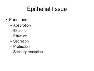
Epithelial Tissue. I. General Features and Considerations. A. Characteristics. Lines internal and external surfaces Single or multiple layers of cells Little or no intercellular space Avascular Polarization Keratin Cell junctions Basement membrane. 1. Protection. 2. Absorption.

E N D
Epithelial Tissue I. General Features and Considerations
A. Characteristics • Lines internal and external surfaces • Single or multiple layers of cells • Little or no intercellular space • Avascular • Polarization • Keratin • Cell junctions • Basement membrane
Epithelial Tissue II. Classification of Epithelia
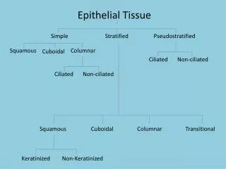
A. Terminology • Classification by number of layers: - simple - stratified • Classification by shape of surface cells: - squamous, cuboidal, or columnar • Classification by structural specializations: - pseudostratified - transitional (urinary) - surface structures
B. Simple epithelia • Simple squamous • Simple cuboidal • Simple columnar
3. Simple columnar epithelium • in profile are rectangular with nuclei usually approximately the same level • most likely to show polarity • often function in absorption, secretion or both • may show extensive surface specializations such as cilia and microvilli • “pseudostratified” columnar - all cells touch the basement membrane (so it’s “simple) but all do not reach the free surface so it looks like it is multilayered
B. Stratified epithelia • Stratified squamous epithelium - Non-keratinized - Keratinized • Stratified columnar (or cuboidal) epithelium
1. Stratified squamous epithelium • usually 5-25 cell layers thick • cuboidal cells on the basement membrane and squamous cells at free surface • found on surfaces subject to injury, wear & tear • non-keratinized - lining inside surfaces - all cells including the surface cells viable - surface cells possess functional nuclei • keratinized - surfaces exposed to external world - surface cells non-viable and do not possess nuclei - surface cells contain almost only keratin (eosinophilic)
2. Stratified columnar (or cuboidal) epithelium • deep cells small, irregularly polyhedral while superficial cells cuboidal or columnar • located at sites of transition from one type of epithelium to another • provides more robust lining than a simple type of epithelium
D. Specialized epithelia • Pseudostratified columnar epithelium • Transitional (urinary epithelium)
1. Pseudostratified Columnar • all cells contact the basement membrane but not all cells reach the free surface • nuclei aligned at two or more levels
2. Transitional Epithelium • form of stratified epithelium that changes in thickness due to the stretch of the organ it lines • when relaxed, the surface cells cuboidal and when distended, the surface cell become more squamous in shape • found in organs of the urinary system
Epithelial Tissue IV. Surface Specializations
A. Microvilli • Large numbers on a cell surface constitute a brush or striated border by light microscopy • 1.0 mm X 0.1 mm evaginations of the luminal plasmalemma
A. Microvilli • Composed of actin filaments, terminal web extends into cytoplasm • Usually covered with a glycocalyx (sugar coat) • Functions - increase surface area for absorption
B. Cilia • Actively motile evaginations of luminal plasmalemma, 2-10 mm long
B. Cilia • Core of longitudinal microtubules called an axoneme (9+2) • Basal bodies • at base of cilia • nine triplet microtubules • Function in transport
Epithelial Tissue V. Basal Lamina
A. Components • Acellular supportive structure that can be up to 100 nm thick • Composed mainly of type IV collagen, laminin, and proteoglycans
C. Functions • Barrier and support • Contains recognition and regulatory factors • Carries a positive charge - thromobogenic