Download

1 / 35
360 likes | 457 Views
EPITHELIAL TISSUE. Definition. Tightly-bound contiguous cells forming sheets covering the body surface or lining the body cavities and their invaginated glands. They are derived from the three embryonic layers (ectoderm, mesoderm and endoderm). Characteristics.

E N D
Definition • Tightly-bound contiguous cells forming sheets covering the body surface or lining the body cavities and their invaginated glands. • They are derived from the three embryonic layers (ectoderm, mesoderm and endoderm).
Characteristics • Cells are tightly bounded together. • Little intercellular spaces and extracellular matrix. • Separated from the underlying connective tissue by basal lamina. • Avascular and it is nourished by diffusion from underlying tissues. • Have a high rate of regeneration.
Functions • Protection. • Trans-cellular transport. • Secretion. • Absorption. • Selective permeability. • Sensation. • Digestion. • Excretion.
Nomenclature • Epidermis for external surface. • Mesothelium for body cavities. • Endothelium for blood vessels. • Neuroepithelium. • Glandular epithelium.
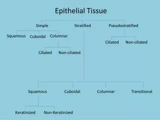
Simple Squamous • Single layer of flat cells with central nuclei. • Function: • Exchange of gases and fluids. • Sites: • Lung alveoli, endothelium, mesothelium, parietal layer of Bowman’s capsule and thin segments of loop of Henle.
Simple Columnar • Single layer of tall cylindrical or hexagonal cells with oval basal nuclei. • Function: • Secretion, absorption, transport. • Sites: • Digestive tract & gall bladder (microvilli), large ducts and female genital ducts & small bronchi (cilia).
Pseudostratified Columnar • Single layer of cells, all rest on basal lamina but not all reach to surface, with nuclei arranged in many rows. • Function: • Protection, secretion, absorption. • Sites: • Respiratory tract ( kinetic cilia). • Epididymis & vas deferens (stereocelia)
Stratified Squamous • Basal cells are cuboidal, next layers are polygonal, the surface cells are flattened. • Function: • protection. • Sites: • Epidermis (keratinized). • Oral cavity, oropharynx, esophagus & vagina (non-keratinized).
Stratified Cuboidal • Two layers of cuboidal cells. • Function: • transport. • Sites: • Duct of sweat glands.
Stratified Columnar • Basal cells are cuboidal, next layers are polygonal, the surface cells are columnar. • Function: • Protection and transport. • Sites: • Conjunctiva of the eye. • Large ducts. • Parts of male urethra
Transitional epithelium • Basal cells are cuboidal or low columnar, next layers are polygonal, the surface cells are columnar, dome-shaped binucleated (in empty bladder), but change to flattened cell in full bladder. • Function: • Protection, transport & adaptation for stretch. • Sites: • Urinary tract (calyces, renal pelvis, ureter, bladder and parts of urethra)
Polarity and cell curface specializations • Epithelial cells have many surface modifications and specializations related to their distinct functionalities. • These specializations occur at all the cell surfaces (apical, lateral and basal surfaces).
Apical specializations • Microvilli. • Cilia. • Flagella. • Cell coat (glycocalyx).
Microvilli • Are small finger-like processes projecting from the free surface of the cell into the lumen. • They increase the surface area of the cell to increase its absorption. • Contain 25-30 actin filaments attached to the apex of the villus in the dense lip. • At the base of the villus, the actin filament are attached to the terminal web. • Microvilli form the brush border of the convoluted tubule of kidney and striated border of the small intestine epithelial cells.
Cilia • Are long motile hair-like processes projecting from the free surface of the cell into the lumen. • Cilia propel the mucus and other structure over the surface of the epithelium by rapid rhythmic oscillations. • Its core is formed of microtubule arranged in axoneme (2 singlets surrounded by 9 doublets). • The cilium is attached to basal body (9 triplets with no singlet).
Flagella • Are similar to cilia, but single and very long. • Present only in the spermatozoa.
Glycocalyx • Glycoprotein attached to the external surface of the cell membrane. • Formed of carbohydrate chain attached to the transmembrane protein of the plasmalemma. • Contains some hydrolytic enzymes and alkaline phosphatase. • They function in protection and cell recognition.
Lateral specializations • Occluding (tight) junction. • Adhering junction. • Desmosome. • Gap junction.
Tight Junctions • Called also zonulae occludentes. • Located between the adjacent plasma membranes. • Are the most apically located junction. • Form belt-like junction at all circumference. • The outer leaflets of the two cell membrane fuse together. • Its function to prevent membrane proteins and water-soluble molecules from passing between the cells.
Adhering Junctions • Called also zonulae adherentes. • It is a junctional complex that lies just basal to the tight junction. • The intercellular space between the outer leaflets is occupied by transmembrane linker protein, which in turn is attached to actin filaments of both adjacent cells. • Function is not only joins the two cell membranes to each other, but also links the cytoskeleton of both cells.
Desmosomes • Called also maculae adherentes. • Are weld-like junctions that help to resist the shearing forces. • Disk-shaped attachment plaques lying opposite each other on the cytoplasmic aspects of plasma membrane of adjacent epithelial cells. • Intermediate filaments are inserted to these plaques, these filaments are responsible for dispersing the shearing forces on the cell.
Gap Junctions • Called also nexus or communicating junctions. • Present in epithelial cells, cardiac and smooth muscles and neurons. • They are tiny pores communcating between two adjacent cells and permit the passage of ions, amino acids, AMP and certain hormones between the cells. • They are under regulation, so they may be opened or closed.
Basal specializations • Basement membrane. • Plasma membrane enfolding. • Hemi-desmosome.
Plasma Membrane Enfolding • Multiple basal enfolding of cell membrane to increase the surface area for transport. • It make many partitions in the basal part of the cells which are occupied by finger-like mitochondria giving the cell striated-appearance. • Present in the ducts of pancreas and salivary glands and are called striated ducts.
Hemi-desmosomes • Attach the basal cell membrane to the underlying basal lamina. • They resemble half desmosomes. • Attachment plaques are present on the cytoplasmic aspect of cell membrane, tonofilaments are attached to these plaques.
Glandular Epithelium • Glands originate from the epithelial surface, penetrate the underlying connective tissue and manufacture a basal lamina around themselves. • The parenchyma represents the secretory units and the duct system, while stroma is the connective tissue between the parenchyma.
Classification • There are many ways for classification of glands according to mode of secretion, type of secretion, way of secretion, number of secretory cells, duct system and shape of the secretory units.
According to presence of ducts • Exocrine gland: means it pours its secretion through duct system to a particular place, e.g. salivary glands. • Endocrine gland: has no ducts and it delivers its secretion directly to the blood stream, e.g. thyroid gland. • Mixocrine gland: has both types, e.g. pancreas.
According to type of secretion • Mucous: sublingual (salivary), Brunner’s glands (in duodenum) and goblets cells. • Serous: parotid salivary gland. • Seromucous: Sunmandibular. • Waxy: ceruminous gland (external ear). • Fatty: sebaceous glands. • Watery: sweat glands. • Milk: mammary glands. • Cellular: testis
According to mode of secretion • Holocrine gland: The whole cell is destroyed and detached, e.g. sebaceous gland. • Merocrine gland: The cell is intact and it secretes through exocytosis of secretory vesicles, e.g. salivary glands. • Apocrine gland: the apical part of the cell is detached, e.g. mammary gland.
According to number of cells • Unicellular gland: one cell only, e.g. goblet cells in the epithelium of mucosa of respiratory tracts and intestine. • Multicellular gland: Many cells and they pour their secretion into the duct system, e.g. salivary glands.
Simple glands • Simple tubular: as in intestine. • Simple coiled tubular: as sweat glands. • Simple acinar. • Simple tubulo-acinar.
Branched glands • Branched tubular: as in stomach. • Branched acinar: as sebaceous glands. • Branched tubulo-acinar: as glands of oral cavity.
Compound glands • Compound tubular: as in kidney and liver. • Compound acinar: as sebaceous glands. • Compound tubulo-acinar: as pancreas and salivary glands.