Download
1 / 10
100 likes | 127 Views
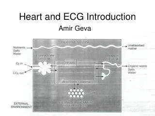
Heart Functions: the ECG and the MEA Feb 15 and 18. How does one calculate heart rate, RR interval, PR interval and ST interval from an ECG? How do these values change pathologically? What leads are used to evaluate the heart during intensive care?

E N D
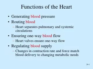
Heart Functions: the ECG and the MEA Feb 15 and 18 • How does one calculate heart rate, RR interval, PR interval and ST interval from an ECG? • How do these values change pathologically? • What leads are used to evaluate the heart during intensive care? • What does the MEA tell us about heart function? • What happens to the MEA in left side or right side hypertrophy?
Lets calculate pulse rate, heart rate, RR interval, PR interval, ST interval from this recording.1) Determine paper speed from X-axis2) Determine distance (mm or cm) between events3) Remember to average at least ECG 3 events (5 is best)4) Re-check math to ensure your units are correct
Lets calculate pulse rate, heart rate, RR interval, PR interval, ST interval from this recording.1) Determine paper speed from X-axis2) Determine distance (mm or cm) between events3) Remember to average at least ECG 3 events (5 is best)4) Re-check math to ensure your units are correct
What are the three standard “LEADS” that folks use to look at heart function? Lead I (RA- and LA+), Lead II (RA- and LL+) and Lead III (LA- and LL+). These form a “perfect” triangle around the heart that sits at the “exact” center of chest. Sort of…
MEAN ELECTRICAL AXIS (MEA) GIVES A MORE ADVANCED CARDIAC DIAGNOSIS OF FUNCTION/PATHOLOGY. • The mean electrical axis is the average direction of depolarization in the heart. • Einthoven’s Triangle: represents an equilateral triangle at the center of chest. (remember the heart is shifted slight to left side) • Dead heart tissue does not depolarize • ECG Changes: Large R-wave becomes a small R-wave • ECG leads can identify exact spot on heart where infarct or clot is located with out cutting you open to look. • MEA if aortic BP is 180/130: • Left Ventricle works harder so it gets larger • MEA shifts to Left Side= Left Shift • MEA if you have emphysema (hard to drive blood through lung): • RightVentricle must work much harder • Pulm arterial pressure 20/480/30 • Right side gets more muscular Mass increasesECG Changes
Normal MEA: 0o to +90o Why Measure MEA: 1) Non-invasive! Do you want them to crack your chest to look around for problems? 2) Its easy! Calculating MEA takes 5 minutes and can be done anywhere anytime. 3) Its cheap! $1 for a few electrodes and 10 cents for paper. However, it is not flawless….you still have to back up what you see with more expensive diagnostics. Now you have a reason to spend the extra cash. What would causes axis deviations? What happens to MEA if you lose 20% of the mass of left ventricle? What happens to MEA if you had a rare right side heart attack and lost 20% of the right ventricle? What happens to size of left ventricle and MEA if aortic valve will not open properly? What happens to MEA if a tumor inside pericardial sac pushes heart to the right side? Easiest Way to Measure MEA to Assess Cardiac Function?
Lets make a Mean Electrical Axis Calculation: Lead I: Right Arm (-) Left Arm (+) Net deflection (mm) =_________ Lead II: Right Arm (-) Left Leg (+) Net Deflection (mm) =_________ Lead III: Left Leg (+) Left Arm (-) Net Deflection (mm) =_________ Estimated MEA= __________degrees What does it mean??______
How to Measure MEA:WHY ECG? Remember: For 75 cents you can record some ECGs and measure an MEA to determine if a heart is “probably” functioning correctly or incorrectly. This is a lot “cheaper” and a lot “less invasive” than immediately cutting a persons chest open for a $75,000 triple cardiac-bypass surgery because they have “chest pain”. Remember Proper Leads: Lead I-Right Arm (-) and Left Arm(+) Lead II: Right Arm (-) and Left Leg (+) Lead III: Left Leg (+) and Left Arm (-) 1) Measure the net amplitude of the QRS complex for leads I, II and III. Net QRS Amplitude: Look at the two largest waves, if the R-amplitude is +9mm and the S-amplitude is -3mm, the net amplitude would be (+9mm) – 3mm = +6mm 2) From the center of each lead line on Einthoven’s Triangle (I, II and III), move that number of mm in the positive or negative direction from the center and place a “mark” at this spot on each lead line. 3) Draw perpendicular lines to each “mark” and circle the place where the three perpendicular lines intersect (or best estimate of the intersection point). 4) Draw a line from the center of the triangle to the center of the intersection. 5) Look and see how many degrees this line is at. This is the average vector taken during the depolarization of the heart that you are evaluating. 6) A Normal heart depolarizes down and towards the left (0o to +90o ). Right Axis Deviation is anything from (+90o to +180o ). Left Axis Deviation is anything from (0o to -90o ). Extreme Right: if MEA is between 180o and -90o recheck the leads, they are probably mixed-up
If you want to become a cardiologist you will use a more complicated 12 lead ECG (Leads I, II, and III, aVR, aVF, aVL, and precordial leads: 1,2,3,4,5, and 6)We won’t try this on any AP212 tests.