Download
1 / 43
430 likes | 666 Views
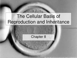
The Cellular Basis of Reproduction and Inheritance. Chapter 8. Asexual Reproduction. Parent cell divides and two ‘daughter cells’ are created Chromosomes and DNA are duplicated 2 daughter cells are identical to each other and to the parent . Sexual Reproduction.

E N D
The Cellular Basis of Reproduction and Inheritance Chapter 8
Asexual Reproduction • Parent cell divides and two ‘daughter cells’ are created • Chromosomes and DNA are duplicated • 2 daughter cells are identical to each other and to the parent
Sexual Reproduction • Offspring produced generally resemble the parent but are not identical to the parents or to each other • Each offspring inherits a unique set of genes from the parent • Highly varied
Cells arise only from preexisting cells • Roles of cell division • Asexual reproduction • Reproduction of an entire single-celled organism • Growth of a multicellular organism • Growth from a fertilized egg into an adult • Repair and replacement of cells in an adult • Sexual reproduction • Sperm and egg production
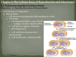
Plasmamembrane Prokaryoticchromosome Cell wall Duplication of chromosomeand separation of copies Continued growth of the cell and movement of copies Division intotwo cells Binary Fission • Prokaryotes reproduce by binary fission, or ‘dividing in half’ • These cells possess a single chromosome, containing genes • The chromosome is replicated • The cell then divides into two cells, a process called binary fission
Eukaryotic Cell Division • A eukaryotic cell has many more genes than a prokaryotic cell • The genes are grouped into multiple chromosomes, found in the nucleus • Chromosomes contain a very long DNA molecule with thousands of genes http://www.botany.org/PlantImages/ImageData.asp?IDN=15-002h&IS=700
Sister chromatids Centromere Chromosomes • Individual chromosomes are only visibleduring cell division • They are packaged as chromatin • Before a cell starts dividing, the chromosomes are duplicated • This process produces sister chromatids
Chromosomeduplication Sister chromatids Centromere Chromosomedistributiontodaughtercells Sister Chromatids • When the cell divides, the sister chromatids separate • Two daughter cells are produced • Each has a complete and identical set of chromosomes http://faculty.uca.edu/~benw/biol1400/notes14.htm
The Cell Cycle • Interphase, where chromosomes duplicate and cell parts are made • The mitotic phase, when cell division occurs • Orderly sequence of events which consists of two major phases
Interphase • Majority of the cells time is spent in interphase • Cells activity is very high • Various metabolic activities • Duplicates chromosomes • Cell parts made, proteins, organelles • Preparation for mitotic division • 3 phases: G1(Gap 1), S (DNA Synthesis), and G2
Mitotic Phase • 2 processes: • Mitosis- nucleus and its contents divide and are evenly distributed to form two daughter nuclei • Cytokinesis- division of the cytoplasm into two daughter cells
The Four Stages of Mitosis • Prophase: • Chromatin coils into distinct chromosomes • Sister chromosomes pair and move towards center of cell • Nucleolus disappears, nuclear envelope fragments • Mitotic spindle begins to form Mitotic Spindle Nuclear membrane Sister Chromosomes
Prometaphase PROMETAPHASE • Spindle microtubules reach chromosomes and attach • Move chromosomes to center • Nuclear envelope disappears Fragments of nuclear envelope Kinetochore Spindle microtubules PROMETAPHASE
Mitotic Spindle Centromere Sister Chromosomes The Four Stages of Mitosis • Metaphase- • The sister chromatids line up in the center of the cell • The spindle fibers form and attach in the center of the chromatids in the centromere
Mitotic Spindle Sister Chromosomes The Four Stages of Mitosis • Anaphase- • The sister chromatids then separate and move to opposite poles of the cell • The spindle fibers from the mitotic spindle pull them apart
Nucleus reforming Cytokinesis The Four Stages of Mitosis • Telophase- • Spindle fibers disintegrate • Chromosomes unwind • Nuclear envelope reforms, nucleus reforms • Cytokinesis splits the cytoplasm • In plants, new cell wall is formed
Cleavagefurrow Cleavagefurrow Contracting ring ofmicrofilaments Daughter cells Cytokinesis • In animals, cytokinesis occurs by cleavage • Ring of microfiliments forms around the circumference of the cell • The ring then contracts • This process pinches the cell apart
Wall of parent cell Cell plate forming Daughter nucleus Cytokinesis • In plants, a membranous cell plate splits the cell in two • Vesicles from the golgi deposit cell wall material into the center • The vesicles then fuse into a cell plate which spans the cell New cell wall Cell wall Vesicles containing cell wall material Daughter cells Cell plate
Cell Growth Factors • Cells must be able to control growth and development in order for an organism to grow normally • In laboratory cultures, most normal cells divide only when attached to a surface • They are anchorage dependent, this keeps cells from dividing in the body while detached • Cells continue dividing until they touch one another • This is called density-dependent inhibition, it keeps cells from overgrowing their organs
Cells anchor to dish surface and divide. When cells have formed a complete single layer, they stop dividing (density- dependent inhibition). If some cells are scraped away, the remaining cells divide to fill the dish with a single layer and then stop (density-dependent inhibition).
Growth Factors • Inadequate supplies of certain growth factor proteins may be the cause of density-dependant inhibition • A growth factor is a protein secreted by certain body cells that stimulate cells in the vicinity to divide • These signals affect critical checkpoints determine whether the cell will go through a complete cycle and divide
Growth factor Plasma membrane Relayproteins G1 checkpoint G1 checkpoint Receptor protein Signal transduction pathway Cell cyclecontrolsystem Controlsystem M checkpoint G2 checkpoint Growth Factors • The binding of growth factors to specific receptors on the plasma membrane is usually necessary for cell division
Cancer Cells • Cancer cells have abnormal cell cycles • They divide excessively and can form abnormal masses called tumors • Radiation and chemotherapy are effective as cancer treatments because they interfere with cell division • Malignant tumors can invade other tissues and may kill the organism
Functions of Mitosis • Growth- • Roots continue to grow in soil • Hair continues to grow on your head • New leaves develop on trees in the fall • Seeds and embryos develop into mature beings
Deadcells Epidermis, the outer layer of the skin Dividingcells Dermis Functions of Mitosis • Cell replacement • Skin replacement • Healing and scarring • Starfish • Asexual Reproduction • Cuttings • Runners • Amoebas • Hydras
Chromosomes Centromere Sister chromatids Homologous Chromosomes • In humans a typical body cell, somatic cell, has 46 chromosomes • 23 matched pairs (4 chromosomes all together), each set of chromosomes has a twin nearly identical in length and centromere position • These matched pairs are called homolgous chromosomes
Homologous Chromosomes • Both carry the genes controlling the same inherited characteristics • Both have the gene controlling the characteristic but they may have a different version of that gene • One has the blue eye version, the other the brown eye version
Sex Chromosomes • Of the 23 pairs: • 22 pairs are autosomes-found in both males and females • The other pair are sex chromosomes that determine gender • Females have a pair of X chromosomes • Males have an X chromosome and a Y chromosome • X and Y chromosomes differ in size and shape
Haploid gametes (n = 23) Egg cell Sperm cell MEIOSIS FERTILIZATION Diploidzygote (2n = 46) Multicellulardiploid adults (2n = 46) Mitosis anddevelopment Gametes • Cells with two sets of chromosomes are said to be diploid • Gametes are the sex cells: sperm and eggs • Gametes are haploid, with only one set of chromosomes • Gametes are formed by a process called meiosis
Meiosis • Meiosis, like mitosis, is preceded by chromosome duplication • However, in meiosis the cell divides twice to form four daughter cells • In the first division, meiosis I, homologous chromosomes are paired • While they are paired, they cross over and exchange genetic information • The homologous pairs are then separated, and two daughter cells are produced
Meiosis reduces the chromosome number from diploid to haploid • Events in the nucleus during meiosis I • Prophase I • Chromosomes coil and become compact • Homologous chromosomes come together as pairs by synapsis • Each pair, with four chromatids, is called a tetrad • Nonsister chromatids exchange genetic material by crossing over
Meiosis reduces the chromosome number from diploid to haploid • Metaphase I • Tetrads align at the cell equator • Anaphase I • Homologous pairs separate and move toward opposite • poles of the cell • Telophase I • Duplicated chromosomes have reached the poles • A nuclear envelope forms around chromosomes in some species • Each nucleus has the haploid number of chromosomes
Meiosis reduces the chromosome number from diploid to haploid • Meiosis II follows meiosis I without chromosome duplication • Each of the two haploid products enters meiosis II • Events in the nucleus during meiosis II • Prophase II • Chromosomes coil and become compact • Metaphase II • Duplicated chromosomes align at the cell equator
Meiosis reduces the chromosome number from diploid to haploid • Anaphase II • Sister chromatids separate and chromosomes move toward opposite poles • Telophase II • Chromosomes have reached the poles of the cell • A nuclear envelope forms around each set of chromosomes • With cytokinesis, four haploid cells are produced
MEIOSIS I: Homologous chromosomes separate INTERPHASE PROPHASE I METAPHASE I ANAPHASE I Centrosomes(withcentriolepairs) Microtubules attached tokinetochore Metaphaseplate Sister chromatidsremain attached Sites of crossing over Spindle Nuclearenvelope Sisterchromatids Tetrad Centromere(with kinetochore) Homologouschromosomes separate Chromatin Meiosis I
MEIOSIS II: Sister chromatids separate TELOPHASE IAND CYTOKINESIS TELOPHASE IIAND CYTOKINESIS PROPHASE II METAPHASE II ANAPHASE II Cleavagefurrow Sister chromatidsseparate Haploiddaughter cellsforming Meiosis II • Meiosis II is essentially the same as mitosis • The sister chromatids of each chromosome separate • The result is four haploid daughter cells
MITOSIS MEIOSIS MEIOSIS I PARENT CELL(before chromosome replication) Site ofcrossing over PROPHASE I Tetrad formedby synapsis of homologous chromosomes PROPHASE Chromosomereplication Chromosomereplication Duplicatedchromosome(two sister chromatids) 2n = 4 Chromosomes align at the metaphase plate Tetradsalign at themetaphase plate METAPHASE I METAPHASE ANAPHASE I TELOPHASE I Homologouschromosomesseparateduringanaphase I;sisterchromatids remain together ANAPHASETELOPHASE Sister chromatidsseparate duringanaphase Haploidn = 2 Daughtercells of meiosis I 2n 2n MEIOSIS II No further chromosomal replication; sister chromatids separate during anaphase II Daughter cellsof mitosis n n n n Daughter cells of meiosis II Mitosis vs Meiosis
Causes of Genetic Variation • 1. Different homologous chromosomes • Each chromosome of a homologous pair comes from a different parent • Each chromosome thus differs at many points from the other member of the pair • The large number of possible arrangements of chromosome pairs at metaphase I of meiosis leads to many different combinations of chromosomes in gametes • Random fertilization also increases variation in offspring
POSSIBILITY 1 POSSIBILITY 2 Two equally probable arrangements of chromosomes at metaphase I Metaphase II Gametes Combination 1 Combination 2 Combination 3 Combination 4 Causes of Genetic Variation
Causes of Genetic Variation • 2. Different versions of the same gene: • The differences between homologous chromosomes are based on the fact that they can carry different versions of a gene at corresponding loci • One chromosome carries one version of a gene, the other carries another
Coat-color genes Eye-color genes C E Brown Black C E C E c e c e c e White Pink Tetrad in parent cell(homologous pair ofduplicated chromosomes) Chromosomes ofthe four gametes Causes of Genetic Variation
3. Crossing Over • Crossing over- the exchange of corresponding segments between two homologous chromosomes • Chiasma- sites of crossing over • During synapsis (when the homologous chromosomes are lined up together) the chromosomes may overlap • When these segments overlap, the overlapping segments may be detached and re-attached to the opposite chromosome
Coat-colorgenes Eye-colorgenes Tetrad(homologous pair ofchromosomes in synapsis) 1 Breakage of homologous chromatids 2 Joining of homologous chromatids Chiasma Separation of homologouschromosomes at anaphase I 3 Tetrad Chaisma Separation of chromatids atanaphase II and completion of meiosis 4 Parental type of chromosome Recombinant chromosome Recombinant chromosome Parental type of chromosome Centromere Crossing Over