Download
1 / 43
430 likes | 447 Views
CHAPTER 8 The Cellular Basis of Reproduction and Inheritance. Modules 8.1 – 8.3. Parents. Reproduction Inheritance. Cell Cycle. Cell Division. Life cycle. Daughter. Parents. Asexual. Reproduction Inheritance. Mitosis. Cell Division. Cell Cycle. Sexual. Meiosis. Daughter.

E N D
CHAPTER 8The Cellular Basis of Reproduction and Inheritance Modules 8.1 – 8.3
Parents Reproduction Inheritance Cell Cycle Cell Division Life cycle Daughter
Parents Asexual Reproduction Inheritance Mitosis Cell Division Cell Cycle Sexual Meiosis Daughter
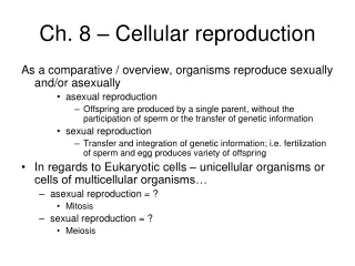
8.1 Like begets like, more or less • Some organisms make exact copies of themselves, asexual reproduction
8.2 Cells arise only from preexisting cells • All cells come from cells • Cellular reproduction is called cell division • Cell division allows an embryo to develop into an adult • It also ensures the continuity of life from one generation to the next
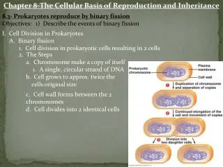
8.3 Prokaryotes reproduce by binary fission • Prokaryotic cells divide asexually • These cells possess a single chromosome, containing genes • The chromosome is replicated • The cell then divides into two cells, a process called binary fission Prokaryotic chromosomes Figure 8.3B
Plasmamembrane Prokaryoticchromosome Cell wall • Binary fission of a prokaryotic cell Duplication of chromosomeand separation of copies Continued growth of the cell and movement of copies Division intotwo cells Figure 8.3A
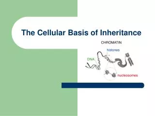
THE EUKARYOTIC CELL CYCLE AND MITOSIS 8.4 The large, complex chromosomes of eukaryotes duplicate with each cell division • A eukaryoticcell has many more genes than a prokaryotic cell • The genes are grouped into multiplechromosomes, found in the nucleus • The chromosomes of this plant cell are stained dark purple Figure 8.4A
Chromosomeduplication • Two daughter cells are produced • Each has a complete and identical set of chromosomes • When the cell divides, the sister chromatids separate Sister chromatids Centromere Chromosomedistributiontodaughtercells Figure 8.4C
8.5 The cell cycle multiplies cells • The cell cycle consists of two major phases: • Interphase, where chromosomes duplicate and cell parts are made • The mitotic phase, when cell division occurs Figure 8.5
8.6 Cell division is a continuum of dynamic changes • Eukaryotic cell division consists of two stages: • Mitosis • Cytokinesis
INTERPHASE PROPHASE Centrosomes(with centriole pairs) Early mitoticspindle Centrosome Fragmentsof nuclearenvelope Kinetochore Chromatin Centrosome Spindlemicrotubules Nucleolus Nuclearenvelope Plasmamembrane Chromosome,consisting of twosister chromatids Figure 8.6
METAPHASE ANAPHASE TELOPHASE AND CYTOKINESIS Cleavagefurrow Nucleolusforming Metaphaseplate Nuclearenvelopeforming Spindle Daughterchromosomes Figure 8.6 (continued)
8.7 Cytokinesis differs for plant and animal cells • In animals, cytokinesis occurs by cleavage • This process pinches the cell apart Cleavagefurrow Cleavagefurrow Contracting ring ofmicrofilaments Figure 8.7A Daughter cells
Cell plateforming Wall ofparent cell Daughternucleus • In plants, a membranous cell plate splits the cell in two Cell wall New cell wall Vesicles containingcell wall material Cell plate Daughtercells Figure 8.7B
8.9 Growth factors signal the cell cycle control system • Proteins within the cell control the cell cycle • Signals affecting critical checkpoints determine whether the cell will go through a complete cycle and divide G1 checkpoint Controlsystem M checkpoint Figure 8.9A G2 checkpoint
8.10 Connection: Growing out of control, cancer cells produce malignant tumors • Cancer cells have abnormal cell cycles • They divide excessively and can form abnormal masses called tumors • Radiation and chemotherapy are effective as cancer treatments because they interfere with cell division
Malignant tumors can invade other tissues and may kill the organism Lymphvessels Tumor Glandulartissue Metastasis 1 A tumor grows from a single cancer cell. 2 Cancer cells invade neighboring tissue. 3 Cancer cells spread through lymph and blood vessels to other parts of the body. Figure 8.10
Parents Asexual Reproduction Inheritance Mitosis Cell Division Cell Cycle Sexual Meiosis Daughter
MEIOSIS AND CROSSING OVER 8.12 Chromosomes are matched in homologous pairs • Somatic cells of each species contain a specific number of chromosomes • Human cells have 46, making up 23 pairs of homologous chromosomes Chromosomes Centromere Sister chromatids Figure 8.12
Haploid gametes (n = 23) Egg cell • The human life cycle Sperm cell MEIOSIS FERTILIZATION Diploidzygote (2n = 46) Multicellulardiploid adults (2n = 46) Mitosis anddevelopment Figure 8.13
MEIOSIS I: Homologous chromosomes separate INTERPHASE PROPHASE I METAPHASE I ANAPHASE I Spindle attached to homologous chromosomes Centrosomes(withcentriolepairs) Metaphaseplate Sister chromatidsremain attached Sites of crossing over Spindle Nuclearenvelope Sisterchromatids Tetrad Centromere Homologouschromosomes separate Chromatin Figure 8.14, part 1
MEIOSIS II: Sister chromatids separate TELOPHASE IAND CYTOKINESIS TELOPHASE IIAND CYTOKINESIS PROPHASE II METAPHASE II ANAPHASE II Cleavagefurrow Sister chromatidsseparate Haploiddaughter cellsforming Figure 8.14, part 2
MITOSIS MEIOSIS PARENT CELL(before chromosome replication) Site ofcrossing over MEIOSIS I PROPHASE I Tetrad formedby synapsis of homologous chromosomes PROPHASE Chromosomereplication Chromosomereplication Duplicatedchromosome(two sister chromatids) 2n = 4 Chromosomes align at the metaphase plate Tetradsalign at themetaphase plate METAPHASE I METAPHASE ANAPHASE I TELOPHASE I ANAPHASETELOPHASE Sister chromatidsseparate duringanaphase Homologouschromosomesseparateduringanaphase I;sisterchromatids remain together Haploidn = 2 Daughtercells of meiosis I 2n 2n No further chromosomal replication; sister chromatids separate during anaphase II MEIOSIS II Daughter cellsof mitosis n n n n Daughter cells of meiosis II Figure 8.15
Meiosis produces genetic variations : Independent orientation of chromosomes POSSIBILITY 1 POSSIBILITY 2 Two equally probable arrangements of chromosomes at metaphase I Metaphase II Gametes Combination 1 Combination 2 Combination 3 Combination 4 Figure 8.16
Coat-color genes Eye-color genes C E Brown Black C E C E c e c e c e White Pink Tetrad in parent cell(homologous pair ofduplicated chromosomes) Chromosomes ofthe four gametes Figure 8.17A, B
Crossing over during prophase I of meiosis Tetrad Chaisma Centromere Figure 8.18A
Coat-colorgenes Eye-colorgenes Tetrad(homologous pair ofchromosomes in synapsis) 1 Breakage of homologous chromatids • How crossing over leads to genetic recombination 2 Joining of homologous chromatids Chiasma Separation of homologouschromosomes at anaphase I 3 Separation of chromatids atanaphase II and completion of meiosis 4 Parental type of chromosome Recombinant chromosome Recombinant chromosome Parental type of chromosome Figure 8.18B Gametes of four genetic types
ALTERATIONS OF CHROMOSOME NUMBER AND STRUCTURE 8.19 A karyotype is a photographic inventory of an individual’s chromosomes • To study human chromosomes microscopically, researchers stain and display them as a karyotype • A karyotype usually shows 22 pairs of autosomes and one pair of sex chromosomes
Fixative Packed red And white blood cells Hypotonic solution Blood culture Stain White Bloodcells Centrifuge • Preparation of a karyotype 3 2 1 Fluid Centromere Sisterchromatids Pair of homologouschromosomes 4 5 Figure 8.19
8.20 Connection: An extra copy of chromosome 21 causes Down syndrome • This karyotype shows three number 21 chromosomes • An extra copy of chromosome 21 causes Down syndrome Figure 8.20A, B
The chance of having a Down syndrome child goes up with maternalage Figure 8.20C
8.21 Accidents during meiosis can alter chromosome number Nondisjunctionin meiosis I • Abnormal chromosome count is a result of nondisjunction • Either homologous pairs fail to separate during meiosis I Normalmeiosis II Gametes n + 1 n + 1 n – 1 n – 1 Number of chromosomes Figure 8.21A
Or sister chromatids fail to separate during meiosis II Normalmeiosis I Nondisjunctionin meiosis II Gametes n + 1 n – 1 n n Number of chromosomes Figure 8.21B
Fertilization after nondisjunction in the mother results in a zygote with an extra chromosome Eggcell n + 1 Zygote2n + 1 Spermcell n (normal) Figure 8.21C
8.22 Connection: Abnormal numbers of sex chromosomes do not usually affect survival • Nondisjunction can also produce gametes with extra or missing sex chromosomes • Unusual numbers of sex chromosomes upset the genetic balance less than an unusual number of autosomes
Poor beardgrowth • A man with Klinefelter syndrome has an extra X chromosome Breastdevelopment Under-developedtestes Figure 8.22A
Characteristicfacialfeatures • A woman with Turner syndrome lacks an X chromosome Web ofskin Constrictionof aorta Poorbreastdevelopment Under-developed ovaries Figure 8.22B
8.23 Connection: Alterations of chromosome structure can cause birth defects and cancer • Chromosome breakage can lead to rearrangements that can produce genetic disorders or cancer • Four types of rearrangement are deletion, duplication, inversion, and translocation
Deletion Duplication Homologouschromosomes Inversion Reciprocaltranslocation Nonhomologouschromosomes Figure 8.23A, B
A chromosomal translocation in the bone marrow is associated with chronic myelogenous leukemia • Chromosomal changes in a somatic cell can cause cancer Chromosome 9 Reciprocaltranslocation Chromosome 22 “Philadelphia chromosome” Figure 8.23C Activated cancer-causing gene
Parents Asexual Reproduction Inheritance Mitosis Cell Division Cell Cycle Sexual Meiosis Daughter