Download
1 / 45
450 likes | 459 Views
The Cellular Basis of Reproduction and Inheritance. Life cycle – the sequence of events that occur between an adult of one generation to an adult of the next. Sexual Reproduction – involves the union of a sperm and an egg, each containing one half of the genetic information.

E N D
The Cellular Basis of Reproduction and Inheritance Life cycle – the sequence of events that occur between an adult of one generation to an adult of the next. Sexual Reproduction – involves the union of a sperm and an egg, each containing one half of the genetic information. Usually involves genetic information from two parents, but always two gametes a sperm and an egg. This type of reproduction requires more energy, but produces valuable differences, or variation, in the offspring.
The Cellular Basis of Reproduction and Inheritance Asexual Reproduction – Is reproduction involving only one parent and no egg or sperm. This process is quick and easy, but produces little or no variation in offspring.
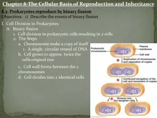
Prokaryotes reproduce by Binary Fission • Binary fission- “dividing in half” • Prokaryotic organisms have only one circular strand of DNA that is their singular chromosome. • This process produces two genetically identical daughter cells.
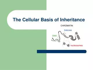
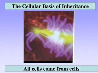
Eukaryotic Chromosomes are more Complex Chromosome – Chroma (colored) soma (body) Chromatin – active threadlike strands of DNA Chromosomes are duplicated long before the cell divides. This forms Sister Chromatids, which are identical chromosomes that are attached at the centromere. They will be separated and pulled into different cells in cell division.
The Cell Cycle Cell Cycle – The cell’s lifecycle. Interphase – The part of the cycle in between cell divisions. G1 – Cell increases in size, produces organelles, and produces proteins. S – Cell copies or synthesizes DNA G2 – Cell produces proteins for division. Mitotic or M phase – Is the division of the nucleus. Cytokinesis – The division of the cell, cytoplasm and organelles.
Functions of Mitosis Growth– mitosis adds new cells to growing tissues. Replacement – Mitosis replaces worn out or damaged cells. Asexual Reproduction Mitosis is used to produce copies of individuals in asexual reproduction.
#37 (in cell div. mitosis song) • #38 (mitosis rap)
Mitosis Prophase– The 1st stage of mitosis • Prepares the nucleus for division, • the nuclear envelope dissolves, • the chromosomes condense becoming visible, • The mitotic spindle forms and attaches to each chromosome at the kinetochore.
Metaphase – the mitotic spindle pulls the chromosomes from each side so that they line up in the Middle on an imaginary line called the Metaphase plate.
Anaphase • Anaphase – The mitotic spindle keeps pulling until the sister chromatids are pulled Apart at the centromeres. This doubles the number of chromosomes in a cell.
Telophase • Telophase – Two new nuclei form • The opposite of prophase • Chromosomes unwind, nuclear envelope reforms
Cytokinesis in Animal Cells • Cytokinesis is the division of the cell’s organelles, cytoplasm and membrane. • In animal cells this starts when a band of microfilaments is formed around the cell and begins to contract producing a cleavage furrow. • The band contracts until the cell is pinched into two new cells.
Cytokinesis in Plant Cells • Plant Cells have a cell wall so they cannot be pinched in half. Instead they form a cell plate made of cellulose that divides the two daughter cells.
Controlling Cell Division • Anchorage Dependence normal cells will not divide unless they are attached to a surface. • Density-Dependent Inhibition Cells stop dividing when they are surrounded by other cells. • Growth Factor Proteins that stimulate cell growth
Cell Cycle Control System • The Red Barriers represent three checkpoints in the cell cycle. • G1 checkpoint – The go ahead signal that starts the S phase. • G2 Checkpoint intiates the M phase or mitosis. • M Checkpoint – indicates that all of the chromosomes are correctly attached to the mitotic spindle.
Mitosis video • #39(mitosis)
Cancer • #40(why don’t we all have cancer)
Cancer Cells are Growing out of Control • A Tumor is an abnormal mass of cells, it can be either benign or malignant. Benign – Always remain in the original site, they do not spread. Malignant – Cells are able to break off and start new tumors where they land, this is called Metastasis, and is what is meant by cancer.
Cancer Cells are Growing out of Control Carcinomas – Cancers of the skin or body covering. Sarcomas – Cancers of the supporting tissues, bones and muscle. Lymphomas and Leukemias – Cancers of the blood forming tissues, spleen, bone marrow or lymph nodes.
Chemotherapy Taxol – prevents the spindle from forming, stopping cell division
HomologousPairs • A somatic cell, or typical body cell, in a human has 46 chromosomes. • These are 23 pairs of homologous chromosomes. • Homologous chromosomes carry the same genes on the same locus, or place on the chromosome. • One chromosome from each of these pairs is inherited from each parent. • 22 pairs of our chromosomes are autosomes, the other pair is the sex chromosomes.
Gametes have half the chromosomes • Cells that contain 23 pairs of homologous chromosomes are referred to as diploid cells. • All human cells are diploid except gametes or sex cells. • Gametes only contain one chromosome from each pair so they are referred to as haploid cells. • Two haploid cells fuse during fertilization producing a diploid zygote. • This new individual will produce gametes by undergoing meiosis to reduce the number of chromosomes by half.
Meiosis overview • #41(how meiosis works)
Meiosis: Diploid to Haploid • In meiosis a cell will divide twice. The first division is meiosis I the second is meiosis II. • Prophase I is similar to that of mitosis with the exception of homologous pairs matching up.
Metaphase I • The homologous pairs are next to each other on the metaphase plate.
Anaphase I • Anaphase I the homologous pairs are pulled apart, leaving sisterchromatids still attached at the centromeres.
Meiosis II • Meiosis II is just like mitosis with only half the chromosomes. • In Metaphase II the chromosomes line up in single file. • Anaphase II the centromeres are broken pulling sister chromatids apart. • Meiosis produces 4 cells from one parent cell. The first division produces genetically different daughter cells, while meiosis II produces 2 identical daughter cells.
Homologous Chromosomes carry different versions of the same genes.
Crossing Over can Increase Variation. • Crossing over occurs when the homologous pairs match up and exchange parts. • This recombination produces new combinations of versions of genes.
Genetic Recombination • This shows how genes on the same chromosome can switch with genes from other chromsomes. • This is called recombination. • Crossing over or recombination occurs in meiosis I. • Crossing over produces new combinations of genes on offspring chromosomes.
Karyotyping • Doctors can take a picture if the nucleus or a karyotype. • The chromosomes are then arranged into homologous pairs and counted. • Extra or missing chromosomes account for many genetic disorders.
Down’s Syndrome: Trisomy 21 • Down’s syndrome is caused by an individual possessing three copies of the 21st chromosome, Trisomy 21. • It result’s in mental retardation that can range from mild to severe, short stature, round face, almond shaped eyes and palm crease. • Down’s syndrome is usually caused by a mistake in egg development, especially in older mothers. • Down’s syndrome occurs in 1 out of 700 children born.
Klinefelter’s XXY • Normal males are XY while an individual with Klinefelter’s syndrome possesses the XX of a female and a Y. • This syndrome results in mental retardation, sterility, breast development, and lack of facial hair.
Turner’s Syndrome XO • Turner’s Syndrome occurs when an individual receives only one X chromosome. • Usually normal intelligence, web of skin on the neck, sterile, constricted aorta, short stature, and poor breast development.
XYY and XXX • XYY“?Jacobs Syndrome”isnot a clearly defined syndrome, though individuals are usually taller than average, lower intelligence, weak musculature and have social problems. • XXX or metafemales have some fertility problems, but are otherwise normal.
ChromosomalAlterations Deletion a fragment is lost and the chromosome reattaches without it. Cri du Chat occurs with a deletion on CH 5 individuals have small heads, severe MR and usually do not survive infancy. Duplication if the lost fragment is reattached in the homologous chromosome that chromosome has two copies of the genes on the fragment.
ChromosomalAlterations Inversion a fragment is reinserted backwards. Translocation occurs when the fragment is reattached in a different chromosome The Philadelphia chromosome occurs when a growth factor gene on CH 9 is moved to CH 22, this leads CML the most common form of leukemia.
Videos #42(mitosis: splitting up CC), #43(Meiosis: where the CC)