Download
1 / 53
530 likes | 710 Views
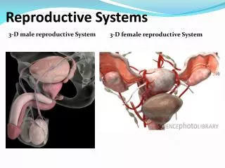
Human Reproductive Systems. Functions of the Reproductive System. Produces and nurtures sex cells Transports sex cells to site of fertilization Secretes hormones vital to the development and maintenance of sexual characteristics Regulates reproductive physiology. Male Reproductive System.

E N D
Functions of the Reproductive System • Produces and nurtures sex cells • Transports sex cells to site of fertilization • Secretes hormones vital to the development and maintenance of sexual characteristics • Regulates reproductive physiology
Testes - Location • Primary sex organs in which sperm cells and male sex hormones are formed • Located within scrotum
Testes - Structure • Surrounded by white fibrous capsule that divides testis into about 250 lobules • Each lobule contains seminiferous tubules • Interstitial cells secrete sex hormones • Tubules unite to form epididymis vas deferens
Spermatogenesis • Process of producing sperm cells • Produced throughout reproductive life • Collect in seminiferous tubules and pass to epididymis to mature
Sperm Structure • Flattened head, cylindrical midpiece, and elongated flagellum • Head contains nucleus (23 chromosomes) • Acrosome • Many mitochondria in midpiece
Epididymis • Tightly coiled, threadlike tube • Connects to ducts of testis • Becomes the vas deferens • Sperm cells are initially nonmotile here
Vas Deferens • Aka ductus deferens • Muscular tube that passes from the testis into the abdominal cavity • Ends behind the urinary bladder • Unites with duct of seminal vesicle to form ejaculatory duct • Ejaculatory duct passes through prostate • Empties into urethra
Seminal Vesicles • Convoluted sac-like structures • Attach to the vas deferens near base of urinary bladder • Secrete slightly alkaline fluid: • Helps regulate pH • Contains fructose to supply sperm with energy • Contains prostaglandins that trigger contractions within female
Bulbourethral Glands • Aka Cowper’s glands • Pair located posterior to the prostate • Produces mucus-like fluid in response to sexual stimulation • Fluid lubricates end of penis
Semen • Fluid conveyed by the male urethra to the outside of the body during ejaculation • Consists of sperm and secretions of the seminal vesicles, prostate, and bulbourethral glands • pH = 7.5 • Volume varies – 2-6 ml • Average sperm count = 120 million / ml • Sperm are nonmotile until they mix with glandular secretions
Scrotum • Pouch of skin that hangs from lower abdominal region behind the penis • Medial septum • Holds the testes
Penis • Conveys urine and semen through the urethra to the outside • Erection • Shaft contains 3 columns of erectile tissue: • Corpora cavernosa • Corpus spogiosum
Penis Structure • Enlarges at distal end to form glans penis • Prepuce • Emission • Ejaculation • Circumcision
Male Sex Hormones • Most changes during puberty are controlled by the hypothalamus (begins around age 10) • Luteinizing hormone (LH) – promotes development of testicular cells that secrete male sex hormones • Follicle-stimulating hormone (FSH) – stimulates cells of seminiferous tubules to respond to testosterone spermatogenesis • Androgens – testosterone; stimulates enlargement of testes and development of secondary sexual characteristics
Male Secondary Sexual Characteristics • Growth of body hair on face, chest, axillary, and pubic regions • Larynx enlarges and vocal cords thicken lower pitch to voice • Skin thickens • Muscular growth increases, shoulders broaden, waist narrows • Bones thicken and strengthen • Increased rate of cellular metabolism and RBC production
Functions of Female Reproductive System • Production and maintenance of ova • Transportation of ova to site of fertilization • Provision of favorable environment for developing offspring • Movement of developed offspring to the outside • Production of female sex hormones
Ovaries - Structure • Solid, ovoid structures • Lie in shallow depressions in lateral wall of pelvic cavity • Inner medulla • Outer cortex
Oogenesis • Primordial follicles formed during prenatal development • Each follicle consists of single primary oocyte • Meiosis begins in primary oocyte and halts until puberty • At puberty, meiosis continues in some primary oocytes • Polar bodies
Follicle Maturation • During maturation of primordial follicle, surrounding follicular cells proliferate, organize into layers, and form fluid-filled cavity around the oocyte. • Follicle cavity presses oocyte to one side, forming “blister” on the surface of the ovary. • 20 primary oocytes may begin maturing, but usually one outgrows the others.
Ovulation • Release of the secondary oocyte and first polar body from the follicle • Hormones cause mature follicle to swell rapidly and its walls weaken • After ovulation, secondary oocyte and 1-2 layers of follicular cells surrounding it are propelled into the uterine tube. • If not fertilized, the oocyte degenerates.
Uterine Tubes • Aka fallopian tubes or oviducts • Open near the ovary and pass medially to the uterus • Infundibulum • Fimbriae
Uterus - Functions • Hollow, muscular organ shaped like an inverted pear • Receives the embryo from the uterine tube and sustains its development • Muscles contract during intercourse to help transport sperm • Size changes greatly during pregnancy
Uterus - Structure • Located in anterior portion of pelvic cavity, above the vagina, and over the bladder • Body – upper 2/3 • Cervix – bottom 1/3; surrounds cervical orifice
Layers of Uterine Wall • Endometrium • Myometrium • Perimetrium • Endometrium and myometrium change extensively during pregnancy • Endometriosis
Vagina • Extends from the uterus to the outside of the body • Conveys uterine secretions • Receives the penis during intercourse • Provides a channel for offspring during birth • Extends up and back into pelvic cavity, posterior to bladder and urethra, anterior to rectum • Hymen
Vulva - Structure • External accessory structure of the female reproductive system • Surrounds openings of urethra and vagina • Labia majora • Mons pubis • Labia minora • Clitoris • Vestibule • Vestibular glands
Female Sex Hormones • Estrogen – produced by the ovaries to stimulate enlargement of accessory organs and external reproductive structures, develops and maintains secondary sexual characteristics: • Development of breasts and ducts of mammary glands • Increased deposition of adipose in breasts, thighs, and buttocks • Increased skin vascularity • Progesterone – produced by ovaries to promote uterine changes during reproductive cycle, affects mammary glands, and helps regulate release of FSH and LH • Androgens – increase hair in pubic and axillary regions; formed by adrenal cortex in females
Female Reproductive Cycle • Aka menstrual cycle • Consists of regular, recurring changes in the uterine lining • Culminates in menses, or menstrual bleeding • Begins around age 13 and continues until middle age • Menopause