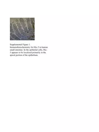
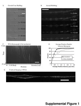
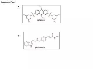
Download
1 / 1
10 likes | 65 Views
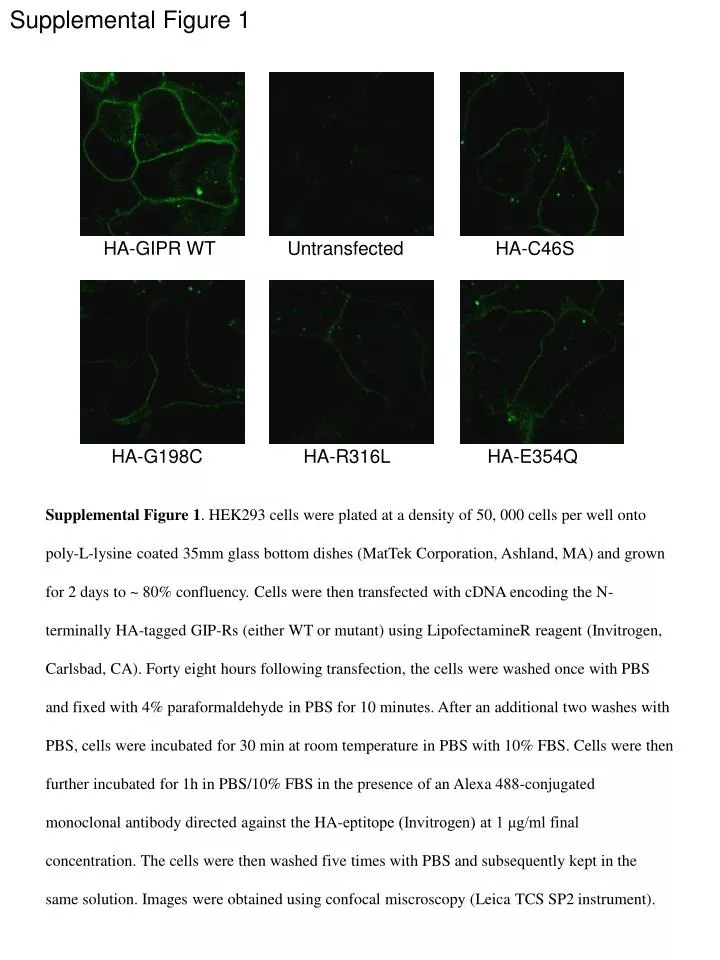
Supplemental Figure 1. HA-GIPR WT. Untransfected. HA-C46S. HA-G198C. HA-R316L. HA-E354Q.

E N D
Supplemental Figure 1 HA-GIPR WT Untransfected HA-C46S HA-G198C HA-R316L HA-E354Q Supplemental Figure 1. HEK293 cells were plated at a density of 50, 000 cells per well onto poly-L-lysine coated 35mm glass bottom dishes (MatTek Corporation, Ashland, MA) and grown for 2 days to ~ 80% confluency. Cells were then transfected with cDNA encoding the N-terminally HA-tagged GIP-Rs (either WT or mutant) using LipofectamineR reagent (Invitrogen, Carlsbad, CA). Forty eight hours following transfection, the cells were washed once with PBS and fixed with 4% paraformaldehyde in PBS for 10 minutes. After an additional two washes with PBS, cells were incubated for 30 min at room temperature in PBS with 10% FBS. Cells were then further incubated for 1h in PBS/10% FBS in the presence of an Alexa 488-conjugated monoclonal antibody directed against the HA-eptitope (Invitrogen) at 1 μg/ml final concentration. The cells were then washed five times with PBS and subsequently kept in the same solution. Images were obtained using confocal miscroscopy (Leica TCS SP2 instrument).