Download
1 / 28
280 likes | 523 Views
The Digestive System By: D. Reis. The digestive tract, also called the gastrointestinal tract (GI) or alimentary canal is a tube that is open at both ends. The 4 Stages of Food Processing. Ingestion – taking in nutrients

E N D
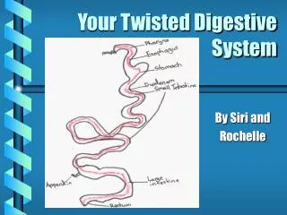
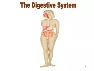
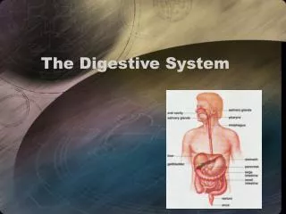
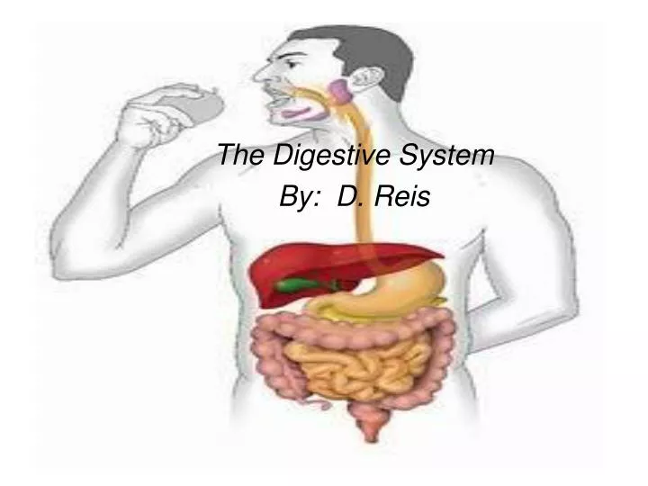
The Digestive System By: D. Reis
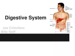
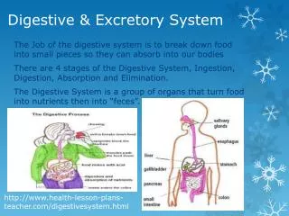
The digestive tract, also called the gastrointestinal tract (GI) or alimentary canal is a tube that is open at both ends.
The 4 Stages of Food Processing • Ingestion – taking in nutrients • Digestion – physically and chemically breaking down food molecules • Absorption – absorbing digested molecules into cells • Egestion – removal of waste food materials
Ingestion and Digestion: Mouth • Incisors (8) – bite • Canine teeth (4) – tear • Premolars – (8) • Molars (12) – crush and grind • Teeth assist in the physical digestion of food.
Tooth Anatomy • Enamel crown – hard substance containing calcium compounds • Dentin – bonelike substance encasing pulp cavity • Pulp cavity – contains nerves and blood vessels
Salivary Glands • The salivary glands secrete saliva, water and amylase. • Amylase– an enzyme released by the salivary glands while chewing. Amlyase breaks down starch (amylose) into maltose subunits.
Pharynx • A bolus enters the pharynx. • Pharynx – dual-purpose passage for food and air • The epiglottis acts like a trap door that prevents food from entering the trachea (windpipe) so that choking does not occur.
Peristalsis • From the pharynx the bolus of food stretches the walls of the esophagus and initiates peristalsis (contractions helping to move food along GI tract) • Peristalsis is independent of gravity
Digestion: Stomach • Sphincters – circular muscles that control movement of food into and out of the stomach • Stomach – contains ridges called rugae that allow it to expand. Cardiac sphincter Pyloric sphincter
Gastric Pits • Gastric pits are openings in the stomach lining which extend into the mucosa as straight and branched tubules, forming gastric glands. Dog stomach to the left
Stomach Secretions • Stomach secretions include : • HCl – helps break down food, kills bacteria that may have been ingested with food and provides an acidic environment for pepsin to function • Mucus – Protects the stomach lining from acidic gastric juices • Pepsin – Enzyme that works at a pH of 2 to help break down protein molecules • Chemical and physical digestion in the stomach changes the bolus of food to partially digested food that contains water and gastric juices with a very low pH called chyme.
Stomach Ulcers • Without mucus, the gastric juice would digest the stomach lining • Damage to the mucus barrier results in gastric ulcers • Most stomach ulcers have been linked to the bacterium H.pylori, which prevents the lining of the stomach from producing mucus
Digestion: Small Intestine • Duodenum – the first 25cm portion of the small intestine where most digestion occurs • Measure up to 7 m in length and 2.5 cm in diameter • In the duodenum chyme is mixed with gall bladder and pancreas contents.
Small Intestine: Digestion and Absorption • Duodenum – food digestion • Jejunum – structure immediately following the duodenum, finalize food digestion • Ileum – structure immediately following the jejunum; absorptive function; linked to large intestine by ileocecal valve
Absorption: Small Intestine • 80% of the absorption of nutrients takes place within the small intestine. • Villi – small fingerlike projections of the mucosa that project into the lumen. • Microvilli – cells making up the lining of each villus • Both increase the surface area of the small intestine for maximum absorption of nutrients into the intestinal wall. • Lacteal – capillary network that allow for diffusion and transport from intestinal cells into the blood.
Mesentery • Anchors the small intestine to the back of the abdominal wall. Blood vessels, nerves, and lymphatics branch through the mesentery to supply the intestine. • Prevents the entanglement of the small intestine.
Small Intestine • The small intestines secretes the following enzymes: • Disaccharidases – breaks down disaccharides into monosaccharides (ex: maltase) • Peptidases – complete the digestion of protein into amino acids
Large Intestine • The large intestine or colon is 1.5 m long and is twice the diameter of the small intestine • Houses bacteria E. coli, which uses waste to synthesize vitamin K and B. • Main function is to reabsorb water (90 % of water is reabsorbed back into the blood) and to remove feces. • Feces consist of cellulose, bacteria and water. Cellulose provides bulk.
Accessory Organs • The liver is the 2nd largest organ in the human body divided into 2 lobes, left and right. • Under the right lobe of the liver is the gallbladder. • The pancreas lies behind the stomach and is about 15cm in length. J
Gall Bladder • The entry of fats into the duodenum stimulates the release of the hormone cholecystokinin (CCK). CCK causes the gall bladder to contract and send bile through the common bile duct to the duodenum. • Bile is synthesized by the liver and stored in the gall bladder. Bile empties into the small intestine where it acts like detergent by physically breaking down large fat globules to smaller fat globules in the small intestine. • The principal components of bile are cholesterol, bile salts, and the pigment bilirubin. Bilirubin is a brownish yellow substance found in bile. It is produced when the liver breaks down old red blood cells. Bilirubin is then removed from the body through the stool and gives stool its normal brown color.
Gallstones • An imbalance between these components of bile cholesterol, bile salts, and bilirubin -- leads to the formation of gallstones. • Gallstones can block the normal flow of bile by lodging in any of the ducts that carry bile thus impairing fat digestion. • Jaundice is a yellowish discoloration of the skin caused by the accumulation of bile in the blood.
The Liver • Catalase found in high concentrations in the liver helps detoxify many harmful substances present in the blood. • Cirrhosis of the liver occurs when damaged liver cells are replaced by connective tissue and fat due to excessive alcohol.
Pancreas • Once chyme enter duodenum the hormone secretin released from the duodenum stimulates the release of Bicarbonate ions (HCO-3)from the pancreas which raises the pH of the chyme from 2.5 – 9, protecting the cells of the duodenum • Because bicarbonate ions increase the alkalinity in the duodenum pepsin becomes inactive. • Lipase – fat digesting enzyme released from the pancreas • Pancreatic amylase – carbohydrate digesting enzyme released from the pancreas • Trypsin and Chymotrypsin – Protein digesting enzyme released from the pancreas