Download
1 / 60
690 likes | 2.01k Views
Digestive System. Joe Pistack MS/ED. Digestive System. Function of Digestive System: Ingestion of food Digestion Absorption of end-products Elimination of waste. Digestive System. The digestive system consist of the following: Mouth Pharynx Esophagus Stomach Small intestine

E N D
Digestive System Joe Pistack MS/ED
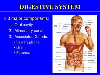
Digestive System • Function of Digestive System: • Ingestion of food • Digestion • Absorption of end-products • Elimination of waste
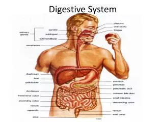
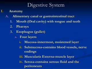
Digestive System • The digestive system consist of the following: • Mouth • Pharynx • Esophagus • Stomach • Small intestine • Large intestine • Rectum • Anus
Digestive System • Accessory organs include: • Salivary glands • Teeth • Liver • Gallbladder • Pancreas • Digestion is process by which food is broken down into smaller particles suitable for digestion • Absorption is the process by which the end products of digestion move across the walls of the digestive tract into blood for distribution throughout the body
Digestion • Two types of digestion: • Mechanical: is the breakdown of large food particles into smaller pieces by physical means • Chemical digestion: is the chemical alteration of food by chemical substances such as digestive enzymes, acid & bile • The end products of digestion are absorbed across the lining of the digestive tract into the blood • Digested nutrients are utilized by the cells of the body • Any food not digested is eliminated from the body as feces • Elimination is the last phase of digestion
Layers of the Digestive Tract • The walls of the digestive tract has 4 layers: • Mucosa: • innermost layer of the tract composed of mucous membrane • Contains cells that secrete mucus, digestive enzymes and hormones • Ducts of exocrine glands empty into the lumen of the digestive tract • Submucosa: • Thick layer of connective tissue that lies next to the mucosa • Contains blood vessels, nerves, glands and lymphatic vessels
Layers of the Digestive Tract • Muscle layer • Third layer of the GI tract • Two layers of smooth muscle consisting of: inner circular layer and outer longitudinal layer • Autonomic nerve fibers innervate the muscle layer • Responsible for mixing movements, contraction and relaxation of the stomach muscles to aid in the mechanical digestion of food • Peristalsis also occurs in the muscle layer which is the rhythmic alternating contraction and relaxation of the muscles that push the food in forward direction through the digestive tract; stimulated by the presence of food • Muscles are also responsible for swallowing and defecation
Layers of the Digestive Tract • Serosa: • Outermost lining of the digestive tract • Extends as the peritoneal membrane • Peritoneal Membranes: • Extension of the serosa • Mesentery and Mesocolon are located behind the digestive organs • Greater and Lesser Omentum are located in front of organs • Form flat and folded structures that: • Help anchor digestive organs • Carry blood and lymph vessels as well nerves to abdominal organs • Restrict the spread of infection in abdominal cavity
Mouth • Mouth • Beginning of digestive tract • AKA oral cavity, buccal cavity • Contains accessory structures: • Teeth—tongue—salivary glands • Teeth • Used to chew food and begin mechanical digestion • Process of chewing food breaking larger particles into smaller is called mastication • Two set of teeth in lifetime: • Deciduous teeth: 20 teeth that appear around 6 months; baby teeth • Permanent teeth: 32 teeth that replace deciduous at 6-12 years
Teeth • Names teeth: • Incisors • Cuspids (canines) • Premolars (bicuspids) • Molars (include wisdom teeth) • Anatomy of the tooth • Crown: above level of the gum (gingiva) covered with hard enamel • Neck: connects crown with root • Root : embedded in jaw bone • Outer surface of root is anchored to periodontal membrane by cementum which hold tooth in place • Most of the tooth contains a bone-like material called dentin • Pulp is connective tissue of tooth that contains nerves and blood vessel within the pulp cavity - extends to root through the root canal
Tongue • Is muscular organ that occupies the floor of the mouth • Two roles: • Facilitates chewing and swallowing by continuously repositioning food in the mouth and assist with swallowing • To taste food • Two structures: • Mucous membrane called frenulum which anchors the tongue to the floor of the mouth • Capillary network that provides sublingual area with rich blood supply
Salivary Glands • Salivary Glands: there are 3 pairs that secrete their contents into the mouth • Parotid glands: largest; lies below and anterior to the ears • Submandibular glands: located in floor of mouth • Sublingual glands: located under the tongue and are the smallest • Secretion of the salivary glands reach mouth by way of tiny ducts • Secrete saliva which is a watery fluid that contains mucus and salivary amylase (ptyalin) a digestive enzyme • 1 liter is secreted daily • Function is to moisten food for swallowing
Hard & Soft Palate • Hard and soft palate form the roof of the mouth • The anterior hard palate separates the oral cavity from the nasal passages • Posterior soft palate separates the oral cavity from the nasopharynx • Soft palate extends toward the back of the oral cavity as the uvula • Uvula is a V-shaped piece of soft tissue that hangs down from the upper back region of the mouth and aids in swallowing • The palatine tonsils are masses of lymphoid tissue located on the sides of the posterior oral cavity and has the role of protection against infection
Pharynx • Pharynx: • AKA throat • Involved in swallowing by reflex action called deglutition • Three parts: • Nasopharynx—oropharynx—laryngopharynx • Only the oropharynx & laryngopharynx are parts of digestive system • The act of swallowing directs food from the pharynx to the esophagus • The epiglottis cover the trachea to prevent aspiration and the opening of the nasophaynx is closed during swallowing
Esophagus • Esophagus: • Tube-like structure that carries the food from the pharynx to the stomach • Approximately 10 inches in length and descends through chest cavity penetrating the diaphragm • Swallowing pushes a bolus of food into the esophagus which stimulates peristaltic activity causing the food to move into the stomach • Glands in the mucosa of the esophagus secrete mucus which lubricates food to facilitate passage
Esophagus • Two sphincters: • Pharyngoesophageal sphincter: located at the top of the esophagus • Gastroesophageal or lower esophageal sphincter (LES): located at the base of the esophagus • Swallowing pushes food past the pharyngoesophageal sphincter • Relaxation of the LES keeps the base of the esophagus open allowing passage of food into the stomach • When contracted LES closes the base of the esophagus preventing reflux or regurgitation
Stomach • Stomach: • Pouch-like organ that lies in the upper left quadrant of the abdominal cavityunder the diaphragm • Performs 5 functions: • Digestion of food • Secretion of gastric juices, digestive enzymes and hydrochloric acid • Secretion of gastric hormones and intrinsic factor (a protein that helps the intestines absorb vitamin B12) • Regulation of the rate and delivery of partially digested food to small intestine • Absorption of small quantities of water and dissolved substances
Stomach • Regions of the stomach: • Fundus - upper portion, closest to the esophagus and lower esophageal sphincter • Body - middle portion of stomach • Pylorus - lower portion of stomach, closest to duodenum • Pyloric canal- continuation of the pylorus • Pyloric sphincter - located at the end of the pyloric canal, regulates the rate of delivery of stomach contents to small intestine • Landmarks of Stomach: • Greater curvature • Lesser curvature
Stomach • Stomach contains rugae which allows for expansion when the stomach is full • When empty the stomach is the size and shape of a sausage • Has the capacity to expand 1 liter • Three layers of stomach muscle: • Longitudinal muscle layer • Circular muscle layer • Oblique muscle layer • The arrangement of the muscle layer allow for churning and mixing of food with gastric juice to create thick paste-like mixture called chyme • Peristalisis moves the propels the food toward the pylorus
Stomach • Nerves of the stomach: • The stomach is innervated by the vagus nerve • Stimulation increases motility and secretion of gastric juices • Glands of the stomach: • The mucus membranes contain gastric glands • The glands are composed of three types of secreting cells: • Mucus cells secrete mucus • Chief cells secrete digestive enzymes • Parietal cells secrete hydrochloric acid and intrinsic factor • Secretion of the gastric glands are called gastric juice • The secretion of thick mucus coats the stomach lining forming a protective barrier preventing the gastric juices from digesting the stomach itself
Small Intestine • Small intestine: • Called small because the diameter is small • Considerable length at 20 feet long • Located in the center lower abdominal cavity • Held in place by the mesentery (extension of peritoneum) • Primary role is chemical digestion and absorption of food • Three parts: • Duodenum • Jejunum • ileum
Small Intestine • Duodenum: • First segment • 10 inches in length • Receives chyme from stomach and secretions from accessory organs (liver, gallbladder and pancreas) • Secretions from mouth, stomach and accessory organs are responsible for digestion of all food • Most digestion and absorption occurs in the duodenum
Small Intestine • Jejunum: • Second segment • 8 feet in length • Some digestion and absorption occurs is first portion of jejunum • Ileum: • Third segment • 12 feet in length • Extends from jejunum to ileocecal valve • Ileocecal valve prevents reflux of contents from the cecum (first part of large intestine) back into the ileum • Ileum is lined with lymphoid tissue called Peyer’s patches which diminishes the bacterial content in the digestive system
Function of the Small Intestine • The walls of the intestine contain circular folds with fingerlike projections called villi • The epithelial cells of each villus form extensions called microvilli • The large number of villi andmicrovilli increase the amount of digested food that can be absorbed • Each villus consists of a layer of epithelial tissue that surrounds a network of blood capillaries and a lymphatic capillaries called a lacteal • The villus absorb the end products of digestion into either the blood capillaries or the lacteal
Function of the Small Intestine • The capillary blood within the villus drains into the hepatic portal vein and into the liver • The end products of carbohydrates and protein digestion first go to the liver for processing before being distributed throughout body • End products of fat digestion enter the lacteal, forming a milky white lymph called chyle which empties directly into the lymph system • The walls of the small intestine also secrete several digestive enzymes and two hormones - secretinandcholecystokinin
Large Intestine • Large intestine • Larger in diameter • 5 feet in length • Extends from the ileocecal valve to the anus • 4 parts: • Cecum • Colon • Rectum • Anal canal
Large Intestine • Cecum: • First part of the large intestine • Located in the right lower quadrant • Ascends on the right side as the ascending colon • Attached to the cecum is the appendix (a structure that contains lymphocytes and is a source of immune cells • Appendix can become inflamed and require surgical removal
Large Intestine • Ascending Colon: • Ascends on the right side of and curves near liver • Transverse Colon: • Crosses the upper abdomen • Descending Colon: • Descends down the left side of the abdomen • Sigmoid Colon: • S-shaped segment • Distal end of large intestine includes • Rectum—anal canal—anus • Anus contains two sphincters : internal & external
Function of Large Intestine • 4 Functions: • Absorption of water and certain electrolytes • Synthesis of certain vitamins by intestinal bacteria (K and some B vitamins) • Temporary storage of feces • Elimination of waste from body • Peristaltic waves move the fecal material from the cecum into the ascending , transverse and descending colon • During the process water is being reabsorbed from the feces, across the intestinal wall into the capillaries which makes feces a semisolid mass
Fuction of the Large Intestine • Bacterial Action: • Escherichia coli (E coli) - part of the normal flora of intestinal bacteria • E coli that is normal in intestinal tract causes serious health risks if in urine or blood • Intestinal bacteria account for 30% of fecal content and almost 100% of the aroma.
Accessory Digestive Organs • Three important organs: • Liver • Gallbladder • Pancreas
Liver • Large reddish-brown organ located in the RUQ in abdomen below the diaphragm and protected by rib cage • Largest gland in the body • 2 lobes - right is larger and left lobe smaller • Separated by a ligament • The ligament secures the liver to the anterior abdominal wall and undersurface of the diaphragm • Liver is surrounded by a tough fibrous membrane called a capsule
Liver Function • Synthesis of bile salts and secretion of bile - bile salts aid in fat digestion and absorption of fat-soluble vitamins • Bile secretion is the main digestive function of the liver • Synthesis of plasma proteins - play role in blood volume and blood coagulation • Storage of glycogen, fat-soluble vitamins (A,D,E,K) and B12 • Detoxification of drugs and other harmful substances which allows for kidneys to excrete
Liver Function- continued • Excretion of bilirubin, cholestrol, drugs and other substances • Metabolism of carbohydrates which in turn regulates blood glucose levels • The liver either stores glucose as glycogen or makes glucose from glycogen dependent upon blood glucose levels • if blood glucose is elevated it stores excess glucose as glycogen • if blood glucose is low the liver converts glycogen to glucose and releases it to blood
Liver - continued • Metabolism of protein - liver can make different amino acids • Converts nitrogen (from ammonia) into urea for excretion by the kidneys • Metabolism of fats - liver breaks down fatty acids, synthesizes cholestrol and phospholipids, and converts excess protein and carbohydrates into fat • Phagocytosis - the kupffer cells (hepatic macrophages) can phagocytose bacteria and other substances within the liver
Blood supply to the Liver • Hepatic portal system: • Is the liver’s unique arrangement of blood vessels • Receives 1.5 liters of blood/minute from the portal vein and hepatic artery • The portal vein drains the blood from all the organs of digestion containing digestive end products • The hepatic artery delivers oxygenated blood to the liver • The blood leaves the liver through the hepatic veins to the inferior vena cava
Liver lobules • Liver lobules: • The liver contains thousand of liver lobules which are the functional unit of the liver • Liver lobules consist of a special arrangement of blood vessels and hepatic cells • There is a central vein with rows of hepatic cells surrounding it • The hepatic cells are bathed with blood that enter the lobule from the hepatic artery and portal vein • Blood from these two blood vessels mixes in the liver in spaces called sinusoids • The hepatic cells extract water and dissolved substances from the sinusoidal blood • The hepatic cells then secrete bile into the tiny canals called canaliculi • These tiny bile canals merge with the canals from other lobules to form larger hepatic bile ducts • Bile exits the liver through the hepatic bile ducts
Bile • Bile: • Green-yellow secretion produced by the liver and stored in the gallbladder • 800-1000 ml is secreted in 24 hours • Composed of water, electrolytes, cholesterol, bile pigments and bile salts • Bile pigments bilirubin and biliverdin are formed from the hemoglobin of old RBC’s • Bile salts are more abundant and aid in digestion of fat and absorption of fat-soluble vitamins and give stool its brownish color
Biliary Tree • Biliary tree: • The ducts that connect the liver, gallbladder, pancreas and duodenum are called the biliary tree • Network of ducts which include the: • Hepatic bile ducts: receives bile from the canaliculi within the liver lobules • Cystic duct merges with the hepatic duct to form the common bile duct • The common bile duct carries both the hepatic ducts and cystic ducts to the duodenum • The base of the common bile duct swells to form the ampulla of Vater (hepatopancreaticampulla) which is the site the main pancreatic duct joins the common bile duct • The sphincter of Oddi (hepatopancreatic sphincter) encircles the base of the ampulla where it enters the duodenum • The sphincter of Oddi is controls the release of bile to the duodenum and is sensitive to nervous, hormonal and pharmacologic control
Gallbladder • Gallbladder: • Pear-shaped sac attached to the underside of the liver • The cystic duct connects the gallbladder with the common bile duct • Bile produced in the liver, flows through the hepatic ducts, cystic ducts and gallbladder • Gallbladder concentrates and stores approx. 1.2 liters/day • The fat in the duodenum stimulates the release of cholecystokinin (hormone) into the blood which travels to the gallbladder causing the smooth muscle of the gallbladder to contract • The contraction of the smooth muscle cause the ejection of bile into the cystic duct then the common bile duct and duodenum
Pancreas • Pancreas • Accessory organ of digestion located just under the stomach • The head of the pancreas rests in the curve of the duodenum and the tail is near the spleen in LUQ of abdomen • The main pancreatic duct travels the length of the pancreas and joins the common bile duct at the ampulla of Vater • The pancreatic duct carries digestive enzymes from the pancreas to the duodenum which is the meeting point for digestion
Pancreas • The pancreas secretes endocrine and exocrine substances • Exocrine substances include: • Pancreatic enzymes, which are the most important digestive enzyme, are secreted by the pancreatic acinar cells in an inactive form and travel through the main pancreatic duct to the duodenum • Alkaline substances, rich in bicarbonate, neutralize the highly acidic chyme coming out of stomach and entering the duodenum • Digestive enzymes in the duodenum work best in an alkaline environment
Pancreas • Endocrine substances: • Secretion of digestive enzymes and bicarbonate are controlled by nervous and hormonal control • The presence of food in the stomach and duodenum is the stimulus for nervous and hormonal response • The presence of chyme in the duodenum stimulates the release of cholecystokinin (CCK) from the duodenal walls • CCK travels in the blood to the pancreas stimulating the release of pancreatic digestive enzymes • The acid in the duodenum stimulates the release of a second hormone, secretin, from the duodenal walls • Secretin travels through the blood to the pancreas stimulating the release of alkaline (bicarbonate) secretions