Download
1 / 41
430 likes | 438 Views
Brain structure and motor functions. Dr. K. Mearow. Terminology refresher. White matter - myelinated fibre tracts Gray matter - areas of neuronal (and glial) cell bodies Tracts - collections of axons subserving similar function or location (CNS) Fasciculus, columns, funiculus

E N D
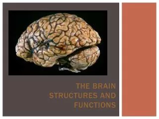
Brain structure and motor functions • Dr. K. Mearow
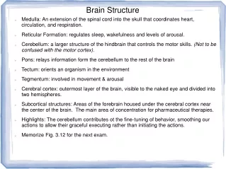
Terminology refresher • White matter - myelinated fibre tracts • Gray matter - areas of neuronal (and glial) cell bodies • Tracts - collections of axons subserving similar function or location (CNS) • Fasciculus, columns, funiculus • Nerves - peripheral axons • Nucleus - collection of neurons subserving similar function (CNS) - eg., caudate nucleus, trigeminal nucleus • Ganglion - collection of neurons in PNS - eg., sensory DRGs, sympathetic ganglia, trigeminal ganglion • Afferent - sensory -information to the CNS • Efferent - motor - information away from CNS towards effector targets
Table 5.3 (1)Page 144 Brain component Cerebral cortex Cerebral cortex Basal nuclei (lateral to thalamus) Basal nuclei Thalamus (medial) Thalamus Hypothalamus Hypothalamus Cerebellum Cerebellum Midbrain Brain stem (midbrain, pons, and medulla) Brain stem Pons Medulla Spinal cord
Major Functions Brain component 1. Sensory perception 2. Voluntary control of movement 3. Language 4. Personality traits 5. Sophisticated mental events, such as thinking memory, decision making, creativity, and self-consciousness Cerebral cortex 1. Inhibition of muscle tone 2. Coordination of slow, sustained movements 3. Suppression of useless patterns of movements Basal nuclei 1. Relay station for all synaptic input 2. Crude awareness of sensation 3. Some degree of consciousness 4. Role in motor control Thalamus 1. Regulation of many homeostatic functions, such as temperature control, thirst, urine output, and food intake 2. Important link between nervous and endocrine systems 3. Extensive involvement with emotion and basic behavioral patterns Hypothalamus 1. Maintenance of balance 2. Enhancement of muscle tone 3. Coordination and planning of skilled voluntary muscle activity Cerebellum 1. Origin of majority of peripheral cranial nerves 2. Cardiovascular, repiratory, and digestive control centers 3. Regulation of muscle reflexes involved with equilibrium and posture 4. Reception and intergration of all synaptic input from spinal cord; arousal and activation of cerebral cortex 5. Role in sleep-wake cycle Brain stem (midbrain, pons, and medulla)
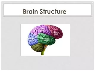
Central sulcus Frontal lobe Parietal lobe Parietooccipital notch Occipital lobe Lateral fissure Preoccipital notch Temporal lobe
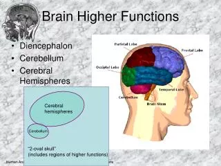
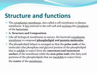
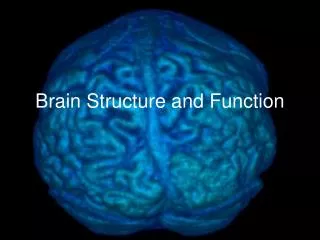
The cerebral cortex • Cerebral Cortex - highly convoluted, outer layer of gray matter. • It covers an inner core of white matter. • The gross structure has gyri (gyrus) and sulci (sulcus) • frontal - voluntary motor activity, speaking ability, and elaboration of thought; stimulation of different areas of its primary motor cortex moves different body regions, again primarily on the opposite side of the body. • parietal - somatosensory processing; each region of its cortex receives somaesthetic and proprioceptive input from a specific body area, primarily from the opposite body side. • temporal- receives sound sensation • occipital- initial processing of visual input
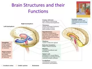
Somatosensory cortex (Somesthetic sensation and proprioception) Supplementary motor area (programming of complex movement) Primary motor cortex (Voluntary movement) Premotor cortex (coordination of complex movements) Posterior parietal cortex (integration of somatosensory and visual input) Central sulcus Prefrontal association cortex (planning for voluntary activity; decision making; personality traits) Parietal lobe Wernicke’s area (speech understanding) Frontal lobe Parietal-temporal-occipital association cortex (integraton of all sensory input- imp in language) Broca’s area (speech formation) Primary auditory cortex Occipital lobe Limbic association cortex (motivation, emotion, memory) Temporal lobe Primary visual cortex
Parietal Lobe - somatosensory cortex • Somesthetic sensation - sensations from the surface of the body - touch, pain, pressure, heat and cold • This info is projected to the somatosensory cortex - site for initial cortical processing and perception of somesthetic and proprioceptive input • Body regions are topographically mapped - sensory homunculus • Sensory cortex - receives information from the opposite side of the body(eg damage on right side results in sensory loss on left side) • Simple awareness of touch, pressure, temp or pain is first detected by the thalamus, but cortex is required for perception - intensity and spatial discrimination • This info is then projected (via fibre tracts) to association cortices for analysis and integration of sensory information - eg., perception of texture, firmness, temp, shape, position, location of an object you are holding)
Figure 5.11 (2)Page 149 Sensory homunculus Left hemisphere Cross-sectional view Temporal lobe
Frontal lobe - motor cortex • Primary motor cortex - voluntary control for muscle movement • Motor cortex on each side controls muscles on the opposite side of the body • Tracts originating in the cortex cross (at level of pyramids) before continuing down spinal cord to terminate on a-motor neurons that directly innervate skeletal muscle • Body regions are represented topographically - motor homunculus • Extent of representation in the motor cortex is proportional to the precision and complexity of motor skills required
Figure 5.12 (2)Page 149 Motor homunculus Left hemisphere Cross-sectional view Temporal lobe
Other cerebral brain regions are important for motor control: • Primary motor cortex does notinitiate voluntary movement • premotor cortex- anterior to the primary motor cortex- acts in response to external cues - must be informed of body’s position in relation to target • supplementary motor cortex - responds to internal cues - plays a preparatory role in programming complex sequences of movement • posterior parietal cortex - It is posterior to the primary somatosensory cortex - informs premotor cortex of position
Occipital Lobe • Primary visual cortex • Receives input from the eyes via optic nerve and optic projections to occipital lobe • Important for coordination of eye movements as well
Temporal lobe • Contains auditory centres that receive sensory fibres from the cochlea of each ear • Also involved in the interpretation and association of auditory and visual information • Temporal lobe contains the hippocampus and the amygdala • Involved in memory
Language • Areas responsible for language ability are found in only 1 hemisphere - usually the left • Language involves the integration of 2 distinct capabilities - expression (speaking ability) and comprehension • Broca’s areais responsible for speaking ability ->frontal lobe - in association with the motor area that controls the muscles necessary for articulation • Wernicke’s areafunctions for language comprehension -> parietal-temporal-occipital association cortex - critical role in understanding both written and spoken language
Language contd • Damage toBroca’s area (expressive aphasia)- failure of word formation, although patient can still understand the spoken and written word • Know what they want to say but cannot express it • Damage toWernicke’s area (receptive aphasia) - loss of understanding of words seen or heard • Can speak fluently, but their words make no sense; cannot attach meaning to words nor choose appropriate words to express thoughts • Concept of words originating in Wernicke’s area must be communicated to Broca’s area - via thearcuate fasciculus • Broca’s area then sends fibre to the motor cortex to control muscles necessary for speech production • Conduction aphasia - damage to thearcuate fasciculus- fluent but nonsensical speech
Facial area of motor cortex Angular gyrus of parietal-temporal-occipital association cortex Broca’s area Wernicke’s area Bundle of interconnecting fibers Visual cortex
Cortical Association areas • prefrontal association cortex- Its functions include planning for voluntary activity, decision-making, creativity, and developing personality traits. • Site of operation of working memory - temporary storage and actively manipulation of information used in reasoning and planning • parietal-temporal-occipitalassociation cortex - It integratessomatic, auditory, and visualsensations from these three lobes • Also involved in connecting Broca and Wernicke’s area • limbic association cortex - It is involved with motivation, emotion, and memory
The cerebral hemispheres - lateralization/dominance • Each cerebral hemisphere receives information from both sides of the body due to connections via the corpus callosum • The left cerebral hemisphere excels in performing logical, analytical, sequential, and verbal tasks • Better at describing facial appearances • The right cerebral hemisphere excels in spatial perception and artistic and musical talents • Better at recognizing faces
The limbic system • Refers to several forebrain structures that function together • Cingulate gyrus • Hippocampus • Amygdala • Septal nuclei • Closed circuit of information flow between the limbic system and the thalamus and hypothalamus • Limbic system and hypothalamus - cooperate in the neural basis ofemotional states
Figure 5.18Page 157 Frontal lobe Cingulate gyrus Fornix Thalamus Hippocampus Temporal lobe Amygdala Hypothalamus Olfactory bulb
Limbic system contd • It plays a key role in emotion and works with the higher cerebral cortex to control behavioral patterns. • Aggression--> lesions of amygdala produce docility, while stimulation results in rage and agression • Fear--> stimulation of amygdala and hypothalamus can produce fear, while ablation results in an absence of fear • Goal-directed behaviour- reward and punishment system- stimulation of certain areas function as a reward, while stimulation of other areas results in a punishment shock • Sex - involved in the regulation of sexual drive and behaviour
Limbic System Highest Level Need Associative Cortex Plan Middle Level Cerebellum Motor Cortex Basal Ganglia Motor Program Spinal Cord Lowest Level Musculo-Skeletal System Movement
Memory • Memory is the storage of acquired knowledge for later recall. • A memory trace is a neural change responsible for retention or storage of knowledge. These traces are present in multiple regions of the brain. • Different systems involved in memory storage and retrieval • Amnesia - results from damage to temporal lobe of cortex, the hippocampus, head of the caudate nucleus (Huntington’s), dorso-medial thalamus (in alcoholics with Korsakoff’s syndrome) • Memory can belong-term (retained for days to years)orshort-term (lasts for seconds to hours) • Short-term involves transient changes in synaptic activity. • Long-term memory involves formation of new, permanent synaptic connections.
Memory contd • Declarative memory- important for remembering facts and events • Two categories - short-term and long-term • People with head trauma may lose memory of recent events but retain older memories • Consolidation of short-term memory into long term memory - function of the medial temporal lobe (hippocampus, amygdala and adjacent areas of cerebral cortex) • Requires gene activation leading to altered protein synthesis and synaptic connections • Amnesia • Retrograde - inability to recall past events - trauma, concussion • Anterograde - inability to store memory in long-term storage for retrieval - damage to medial temporal lobe
Memory contd • Thehippocampusplays a vital role inshort-term memory. It works with other brain regions for declarative memories. • Alzheimer’s disease - damage is seen in hippocampus and cortical areas • Theprefrontal cortexfunctions forworking memory • Temporary storage site, but also responsible for executive functions involving manipulation and integration of the info for planning, problem-solving and organizing activities • Thecerebellumfunctions forprocedural memories.
The basal nucleii (ganglia) have an inhibitory role in motor control
Basal Ganglia • Act by modifying ongoing activity in motor pathways • Inhibit muscle tone (proper tone - balance of excitatory and inhibitory inputs to motor neurons that innervate skeletal muscle • Select and maintain purposeful motor activity while suppressing unwanted patterns of movement • Monitor and coordinate slow and sustained contractions, especially those related to posture and support
Huntington’s disease- degeneration of thecaudate nucleus • Chorea - rapid, uncontrolled jerky movements • Parkinson’s disease - damage to basal ganglia neurons and a deficiency indopamine • Increased muscle tone or rigidity • Resting tremors (eg unwanted movements • Slowness in initiating and carrying out motor behaviours
The Thalamus • Thalamus is a relay station • It is also a synaptic integrating center for processing sensory input on its way to the cerebral cortex. • It also serves to integrate information important for motor control • Receives sensory information from different areas of the body • Information is processed by specific thalamic nuclei
Hypothalamus - homeostatic control • body temperature • thirst and urine production • food intake • anterior pituitary hormone secretion • production of posterior pituitary hormones • uterine contractions and milk ejection • serves as an ANS coordinating center • plays a role in emotional and behavioral patterns
Cerebellum Regulation of muscle tone, coordination of skilled voluntary movement Planning and initiation of voluntary activity Vestibulocerebellum Maintenance of balance, control of eye movements Spinocerebellum Cerebrocerebelum
Reticular activating system Cerebellum Visual impulses Reticular formation Brain stem Auditory impulses Spinal cord Ascending sensory tracts Descending motor tracts
The brain stem --> the medulla, pons, and midbrain. • Vital link between the spinal cord and higher brain regions - its size belies its importance • Does the same basic sensory and motor functions for the head that the spinal cord does for the rest of the body • Control centers for heart and blood vessel function, respiration, digestive activities • Plays a role in modulating the sense of pain • Plays a role in regulating muscle reflexes involved in equilibrium and posture
Midbrain • several characteristic structures - superior (visual) and inferior (auditory) colliculi • Cerebral peduncles - projection fibres from motor neurons in motor cortex - go to spinal cord, pons and medulla • Substantia nigra • Red nucleus • Sensory fibres ascending to thalamus • Fibres from cerebellum
Pons • Area that contains numerous fibre bundles • Pontine nuclei • Contains cranial nerve nuclei • Cerebellar fibres • Continuation of the fibre tracts that arose in cortex
Medulla • Characteristic structural feature --> • pyramids - area of decussation of projection fibres from cortical motor neurons • More cranial nerve nuclei • Sensory projection fibres from dorsal columns terminate in medulla nuclei
Reticular formation • Core of the brainstem • Receives and integrates the information carried by most of the sensory, motor and visceral pathways that pass through the brainstem • Info is used in various reflexes - circulatory and respiratory reflexes, coughing, swallowing • Plays a role in modulating sensitivity of spinal reflexes and regulating transmission of sensory info (esp pain) into ascending pathways • Ascending fibres carry signals to arouse and activate the cerebral cortex • RAS - controls the overall degree of cortical alertness; important in the ability to direct attention
Cranial Nerves • 12 cranial nerves • Serve general motor and sensory function • Also ‘special’ afferent and efferent (visceral innervation)
Cranial nerves = Motor fibers = Sensory fibers I-Olfactory nerve Retina Mucosa of nasal cavity IV - Trochlear nerve III- Oculomotor nerve II- Optic nerve Termination of fibers of olfactory nerve VI -Abducens nerve Motor— muscles of mastication Olfactory bulb Lateral rectus Sensory—face and head V- Trigeminal nerve Motor—muscles of face and scalp; salivary and tear glands Sensory— taste buds on anterior tongue VII - Facial nerve
Motor—muscles of pharynx; parotid gland Sensory—taste buds on posterior tongue; receptors in pharynx and carotid sinus = Motor fibers = Sensory fibers Vestibular branch VIII - Vestibulocochlear nerve Cochlear branch Cochlea, vestibule, and semicircular canals of inner ear IX - Glossopharyngeal nerve XII - Hypoglossal nerve X - Vagus nerve XI - Accessory nerve Motor—muscles of pharynx and larynx; thoracic and abdominal organs Tongue muscles Muscles of larynx, pharynx, soft palate, shoulder, and neck Sensory—taste buds on tongue and pharynx; thoracic and abdominal organs