Download
1 / 6
70 likes | 120 Views
KIDNEY RLO 1 – anatomy. Page 1. Kidney gross anatomy. Inferior vena cava.

E N D
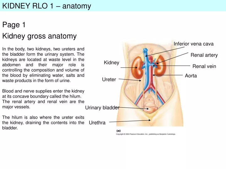
KIDNEY RLO 1 – anatomy Page 1 Kidney gross anatomy Inferior vena cava In the body, two kidneys, two ureters and the bladder form the urinary system. The kidneys are located at waste level in the abdomen and their major role is controlling the composition and volume of the blood by eliminating water, salts and waste products in the form of urine. Blood and nerve supplies enter the kidney at its concave boundary called the hilum. The renal artery and renal vein are the major vessels. The hilum is also where the ureter exits the kidney, draining the contents into the bladder. Renal artery Kidney Renal vein Aorta Ureter Urinary bladder Urethra
KIDNEY RLO 1 – anatomy Page 2 Renal capsule Kidney internal structure Cortex The kidney is surrounded by a fibrous capsule called the renal capsule. Internally there is an outer cortex layer, and inner medulla which comprises of the medullary pyramids which are striped in appearance. This striped feature is due to the position of the tubes and collecting ducts associated with the nephron, the functional unit of the kidney (see page 3). Urine formed in the tubules and collecting ducts of the kidney drain into the renal pelvis, and exits the kidney via the ureter. Renal pelvis Medullary pyramid Ureter Fig 2. Vertical section of the kidney
KIDNEY RLO 1 – anatomy Page 3 The nephron Distal convoluted tubule Bowman’s capsule The nephron is the basic functional unit of the kidney, and there are around 1 million per kidney. The nephron is a tube with different regions specialised in different functions. The nephron begins with a cup-like structure called Bowman’s capsule which opens into a coiled region of tube called proximal convoluted tubule. This tubule thins and straightens into the loop of Henle, which then forms another coiled region called the distal convoluted tubule. The distal tubule empties into the collecting duct. The long Loop of Henle extends down into the renal medulla of the kidney. Proximal convoluted tubule Renal cortex Renal medulla Loop of Henle Collecting duct Fig 3. The nephron
KIDNEY RLO 1 – anatomy Page 4 Cortical Nephron Types of nephron Juxtamedullary Nephron There are two types of nephron. The juxtamedullary nephron has a long loop of Henle which penetrates into the medulla. The cortical nephron which is shorter and remains within the renal cortex. Renal cortex Renal medulla Fig 4. Types of nephron
KIDNEY RLO 1 – anatomy Page 5 Glomerulus Efferent arteriole Afferent arteriole Blood supply to the nephron In order to perform its function of removing waste products from the blood, and regulating water and electrolyte balance, the nephrons have a rich blood supply. The renal artery branches into high-pressure afferent arterioles which form a knot of capillaries called the glomerulus, which sit within the Bowman’s capsule. The glomeruls drains into the efferent arteriole (Efferent = Exit) which entwines the kidney tubules and forms large loops parallel to the loop of Henle, called the vasa recta. The blood vessels re-join and exit the kidney in the renal vein. Vasa recta Fig 5. Blood supply to the nephron
KIDNEY RLO 1 – anatomy Page 6 B Passage of Urine A Urine formed in each nephron drains down the collecting ducts (A). The collecting ducts empty into the renal pelvis (B). Urine exits via the ureter, which joins each kidney to the bladder (C). From the bladder, urine is voluntarily eliminated through a small tube called the urethra, in the process of micturation (C). Ureter Bladder C Urethra